
A thyroid ultrasound is one of the simplest imaging tests there is: no needles, no radiation, no fasting, and usually about half an hour from start to finish. Even so, people often arrive unsure why it was suggested, what the sonographer is looking at, and what the words on the report will mean. This guide walks through the reasons a doctor may recommend a thyroid ultrasound, what the exam is like, what is measured, what a typical report contains, where TI-RADS fits in, and, just as important, what an ultrasound cannot tell anyone.
Here is our role, stated plainly: we never diagnose. Our sonographer performs the scan, a board-certified radiologist reads it and writes the report, and your own doctor interprets that report for you, together with your exam, your lab results and your history. Nothing in this article tells you what your own result means; it is background to help you have a better conversation with your physician.
What the thyroid is and why it is easy to image
The thyroid is a butterfly-shaped gland in the front of the lower neck, wrapped around the windpipe. It has a right lobe and a left lobe joined by a thin bridge called the isthmus. Because it sits just under the skin, high-frequency ultrasound can show it in fine detail. Ultrasound uses sound waves rather than ionizing radiation, and Cleveland Clinic describes the test as quick and painless with no known risks.
Why doctors recommend a thyroid ultrasound
According to MedlinePlus, RadiologyInfo and Cleveland Clinic, common reasons include:
- A lump or swelling in the neck. RadiologyInfo notes that ultrasound can help determine whether a lump is arising from the thyroid or from a nearby structure. Our post on neck lumps and swelling covers this in more depth.
- A finding on a physical exam, such as a nodule, an enlarged or irregular gland (goiter), or abnormal lymph nodes near the thyroid (MedlinePlus).
- Abnormal thyroid blood tests or symptoms that suggest a thyroid problem (Cleveland Clinic).
- A nodule seen by chance on a CT, MRI or other scan done for another reason.
- Follow-up of a known nodule, to see whether it has changed in size or appearance over time (RadiologyInfo).
If you are not sure why an ultrasound was suggested for you, ask your doctor what question they hope it will answer. That makes the report much easier to understand later.
Screening without symptoms: what the USPSTF says
The U.S. Preventive Services Task Force (USPSTF) recommends against screening for thyroid cancer in adults without symptoms. This is a grade D recommendation, issued in May 2017, and it covers screening by neck examination and by ultrasound. In USPSTF terms, a D grade means there is moderate or high certainty that the service has no net benefit or that its harms outweigh its benefits. A key concern the Task Force describes is overdiagnosis: finding and treating small cancers that would never have caused problems.
The USPSTF statement does not apply to everyone. It does not cover people with symptoms such as hoarseness, neck pain or trouble swallowing; people with a lump, swelling or asymmetry in the neck; people with a history of exposure to ionizing radiation (for example, radiation treatment as a child or fallout exposure); people with an inherited syndrome linked to thyroid cancer; or people with a parent, sibling or child who had thyroid cancer. Whether a thyroid ultrasound makes sense for you is a decision to make with your doctor, who can weigh your history and risk factors.
Before the exam: no prep, open neckline
- No prep. There is no fasting and no need to drink water. You can eat, drink and take your usual medications.
- Wear an open neckline. A V-neck, scoop neck or button-up shirt works well. Avoid turtlenecks and high collars.
- Leave necklaces and neck jewelry at home, or plan to remove them.
- Bring any prior thyroid imaging reports and a list of recent thyroid blood test results if you have them, so you can share them with your doctor alongside the new report.
During the exam: what it feels like
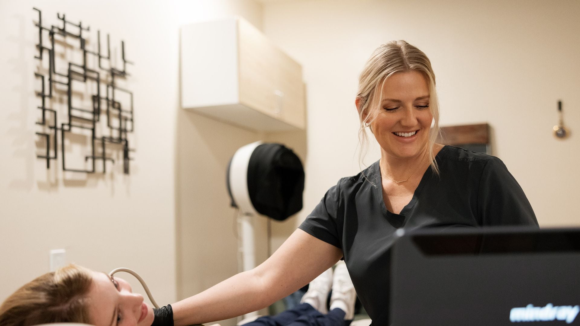
You lie on your back. MedlinePlus describes the neck resting on a pillow or other soft support and stretched slightly; often a small pillow or rolled towel under the shoulders lets the head tilt back so the front of the neck is extended and easier to reach. The sonographer applies a water-based gel, which may feel cool, and moves a small handheld transducer over the front and sides of the neck, taking images from several angles. MedlinePlus says you should feel very little discomfort; some people notice light pressure.
Cleveland Clinic says the test takes about 30 minutes from start to finish, though this varies; the scanning itself is often shorter, and it may take longer when there are several nodules to measure. Afterward the gel is wiped off, and you can go straight back to your day. Our sonographer will not give results during the visit, which is standard practice; our post on why the sonographer won’t tell you what they see explains why.
What a thyroid ultrasound measures
| What is looked at | What is typically recorded |
|---|---|
| Gland size | Length, width and depth of each lobe, and the thickness of the isthmus |
| Overall appearance (echotexture) | Whether the tissue looks even or uneven (heterogeneous), which can be seen with inflammation such as thyroiditis |
| Blood flow | Often assessed with color Doppler, describing flow within the gland or a nodule |
| Nodules | Location, size in three dimensions, and features such as composition, echogenicity, shape, margins and any bright spots (echogenic foci) |
| Cysts | Fluid-filled areas and their size |
| Nearby lymph nodes | Whether any neck lymph nodes stand out by size or appearance |
Cleveland Clinic lists thyroid size and shape, growths or nodules, inflammation and enlargement among the things the test can show. Thyroid nodules are very common, and the ACR notes that many of them are benign. A measurement or description on its own does not tell anyone what it means for a particular person; your physician will put it in context with your history, labs and symptoms.
What the radiologist report includes
A board-certified radiologist reads every study, and the report is ready within 24 hours. It goes to you and to the doctor you name in the booking form. Reports vary, but a thyroid ultrasound report usually follows this outline:
- Indication: the reason for the exam, such as “palpable nodule” or “follow-up.”
- Comparison: whether prior imaging was available. Sharing earlier reports helps the radiologist describe change over time.
- Findings: lobe and isthmus measurements, echotexture, blood flow, each nodule described by location, size and features, and any lymph nodes of note.
- Impression: the radiologist’s summary, which may include a TI-RADS category for each nodule and a suggestion such as follow-up imaging or further evaluation, for your doctor to consider.
The impression is written for your physician, who decides what happens next. Our guides to what an ultrasound report means and common report words explain the vocabulary, and your doctor will explain what it means for you.
TI-RADS in one paragraph
Many radiologists use ACR TI-RADS, the American College of Radiology’s Thyroid Imaging Reporting and Data System, published in a 2017 white paper. It gives points for five ultrasound features of a nodule (composition, echogenicity, shape, margin and echogenic foci) and adds them up into a level from TR1 (“benign”) through TR2 (“not suspicious”), TR3 (“mildly suspicious”), TR4 (“moderately suspicious”) and TR5 (“highly suspicious”). Each level is paired with size thresholds that help a physician decide between no follow-up, repeat ultrasound or fine-needle aspiration; for example, TR5 nodules reach the biopsy threshold at 1 cm, while TR3 nodules reach it at 2.5 cm. The ACR describes the goal as reducing unnecessary biopsies of benign nodules. A TI-RADS level is a risk category, not a diagnosis, and your doctor will interpret it with everything else they know. For the full scoring, see thyroid nodules and TI-RADS explained.
What a thyroid ultrasound can’t tell
- It is not a biopsy. RadiologyInfo states that the radiologist cannot distinguish between benign and malignant lumps with complete certainty, and Cleveland Clinic notes that ultrasound can’t diagnose cancer. When a closer look is needed, a physician may recommend a fine-needle biopsy, which examines cells under a microscope, or a repeat ultrasound after a few months. We do not perform biopsies; your doctor can tell you where they are done.
- It does not measure thyroid function. RadiologyInfo is explicit that function can’t be determined by ultrasound; that takes blood tests (such as TSH) or a radioactive iodine uptake test. We do not draw blood; thyroid bloodwork is usually done at labs such as Quest or Labcorp, through your doctor.
- It does not explain symptoms on its own. Fatigue, weight change, palpitations and many other symptoms have many possible causes. Your doctor combines the ultrasound with your exam and labs.
Questions to ask your doctor
- What question is this ultrasound meant to answer for me?
- Do I need thyroid blood tests as well, and where should I get them?
- If the report lists a nodule, what does its size and TI-RADS level mean in my situation?
- Is follow-up imaging recommended, and when?
- Would a biopsy or a specialist referral help, and why or why not?
- Should I bring my prior imaging to compare?
When to go to the ER or call 911
An outpatient ultrasound is not emergency care. Go to the emergency room, or call 911, for:
- Trouble breathing or noisy, high-pitched breathing
- Trouble swallowing, including being unable to swallow saliva
- Neck swelling that grows quickly over hours or a few days
- A very fast or irregular heartbeat with chest pain, confusion, high fever or fainting
For a new lump that is not growing quickly and is not affecting breathing or swallowing, contact your doctor promptly; they can decide how soon you should be seen and which tests make sense.
Getting your scan with us
You can book a thyroid ultrasound directly at our booking page; no referral is needed. The booking form asks for the name of the doctor who should receive the report (type “None” if you don’t have one) and offers an optional referral upload. Visits are at our clinic, or we bring the scan to you across our California service areas with a portable diagnostic system. You pay a flat rate when you book, with no facility fee. HSA/FSA cards are accepted; we don’t bill insurance, and a superbill is available. See our thyroid ultrasound page for details, and our metabolic screening post if your doctor has also suggested an abdominal scan.
Sources
- U.S. Preventive Services Task Force: Thyroid Cancer: Screening (2017)
- RadiologyInfo (ACR/RSNA): Ultrasound, Thyroid
- Cleveland Clinic: Thyroid Ultrasound
- MedlinePlus: Thyroid ultrasound
- American College of Radiology: ACR TI-RADS
- Tessler FN, et al. ACR TI-RADS: White Paper of the ACR TI-RADS Committee. J Am Coll Radiol. 2017.
Frequently asked questions
How do I prepare for a thyroid ultrasound?
No prep is needed. You can eat, drink and take your usual medications. Wear an open neckline and leave necklaces at home.
How long does a thyroid ultrasound take?
Cleveland Clinic says about 30 minutes from start to finish, though it varies. It may take longer when there are several nodules to measure.
Does the USPSTF recommend thyroid ultrasound screening?
No. The USPSTF recommends against screening for thyroid cancer in adults without symptoms (grade D, 2017), by neck exam or ultrasound. It does not apply to people with symptoms, a neck lump, radiation exposure, an inherited syndrome or a close relative with thyroid cancer. Talk with your doctor about whether a scan makes sense for you.
Can a thyroid ultrasound tell if a nodule is cancer?
No. Ultrasound describes a nodule’s size and features, and a radiologist may assign a TI-RADS risk level, but it cannot tell benign from malignant with certainty. Your doctor may recommend follow-up imaging or a biopsy.
Does a thyroid ultrasound check thyroid function?
No. Thyroid function is measured with blood tests such as TSH. We do not draw blood; your doctor can order labs at places such as Quest or Labcorp.
Who gets the report, and when?
A board-certified radiologist reads every study, and the report is ready within 24 hours. It goes to you and to the doctor you name when booking. Your doctor interprets it for you.
This article is general health information, not medical advice. It does not replace a conversation with your own physician, who knows your history and can interpret results in context. If you have symptoms that worry you, contact your doctor; if you think you are having a medical emergency, call 911.