
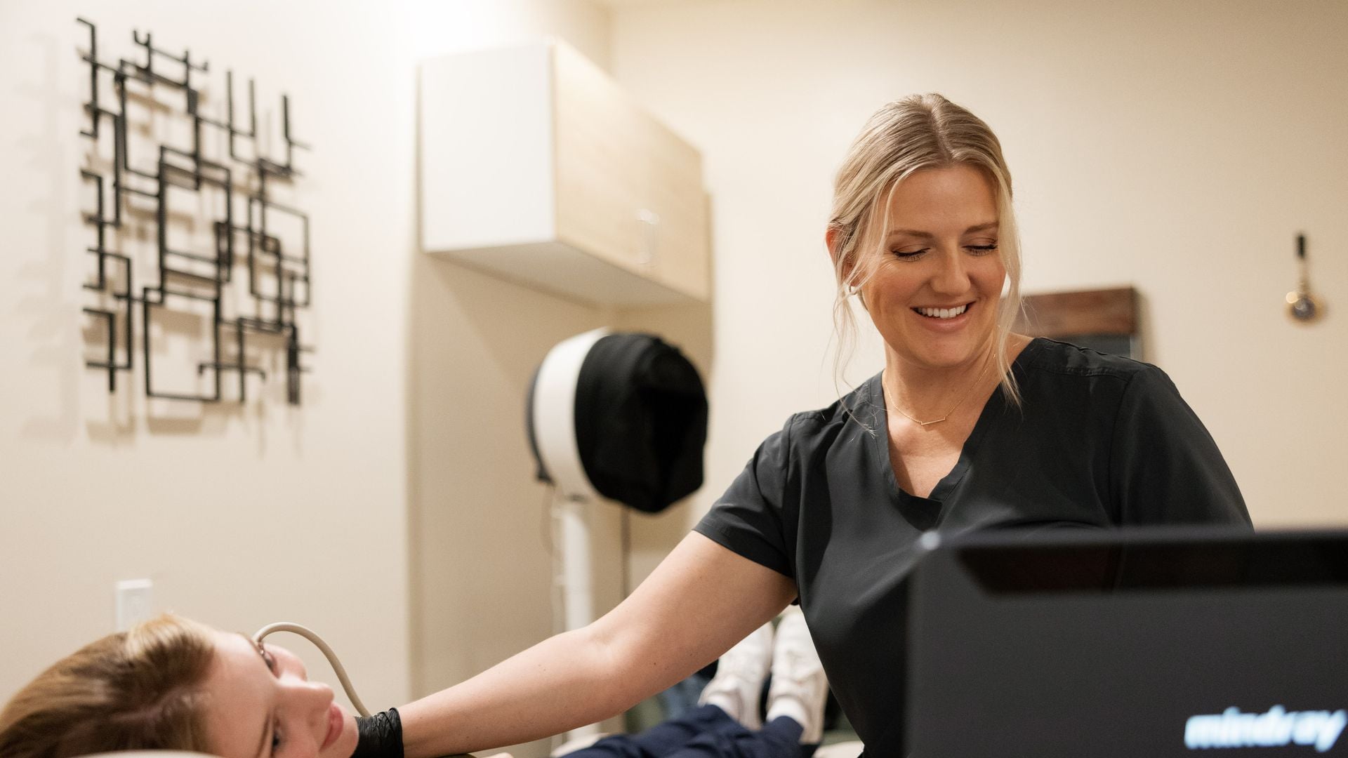
“Vascular ultrasound” is an umbrella term. It covers several different scans of arteries and veins, and each one answers a different question. A carotid duplex looks at the arteries in the neck that carry blood to the brain. An abdominal aortic aneurysm (AAA) scan measures the main artery in the belly. A deep vein thrombosis (DVT) study looks for blood clots in the deep veins of the legs or arms. They all use the same basic tool, sound waves plus Doppler, but they are performed differently, they need different preparation, and the national screening recommendations for them are not the same.
This guide explains what each scan is, what it looks at, what the U.S. Preventive Services Task Force (USPSTF) says about using it in people without symptoms, how to prepare, and which vascular studies we do not offer. It is written to help you have a better conversation with your own doctor about which scan, if any, makes sense for you. It does not tell you which scan you need, and it does not tell you what any result means for you.
Our roles are the same for every study: our sonographer performs the scan, a board-certified radiologist reads the images and writes the report, and your own physician interprets that report for you in light of your history, blood pressure, cholesterol, labs and symptoms. We never diagnose.
Emergencies first: when to call 911 instead of booking a scan
A scheduled ultrasound is never the right response to a vascular emergency. We are not an emergency service. If you or someone near you has any of the symptoms below, call 911 right away, even if the symptoms ease or come and go.
Possible stroke: think BE FAST. Cleveland Clinic describes the signs this way:
| Letter | What to watch for |
|---|---|
| B: Balance | A sudden loss of balance |
| E: Eyes | Sudden vision loss or changes in one or both eyes |
| F: Face | A droop on one or both sides of the face; ask the person to smile |
| A: Arms | When both arms are lifted, one sags or drops in a way it usually doesn’t |
| S: Speech | Slurred speech or trouble choosing the right words |
| T: Time | Call 911 immediately and note when the symptoms began |
Possible blood clot in the lungs (pulmonary embolism). The CDC lists difficulty breathing, a faster than normal or irregular heartbeat, chest pain or discomfort that usually gets worse with a deep breath or coughing, coughing up blood, and very low blood pressure, lightheadedness or fainting. The CDC says to seek medical help immediately. A person can have a pulmonary embolism without any leg symptoms first.
Possible ruptured or leaking aortic aneurysm. Mayo Clinic lists sudden, very bad and long-lasting belly or back pain that may feel like ripping or tearing, along with low blood pressure and a fast pulse. Feeling faint with that kind of pain is a reason to call 911 now.
New swelling, pain, warmth or redness in one leg or arm is not always a 911 situation, but it is a reason to contact your doctor the same day, because the CDC says these can be signs of a deep vein clot. If any of the breathing or chest symptoms above appear, treat it as an emergency.
What Doppler is, in plain language
A regular ultrasound sends sound waves into the body and turns the returning echoes into a black-and-white picture of structures, such as an artery wall. Doppler adds information about movement. Cleveland Clinic explains that the sound waves bounce off moving blood cells and return to the probe, and the change in the sound’s pitch lets the computer estimate which direction blood is flowing and how fast. That is why you may hear a whooshing or swishing sound from the machine during a vascular scan. It is normal and it is part of the exam.
Several Doppler modes are commonly used together:
- Color Doppler: overlays color on the picture to show the direction of flow.
- Spectral Doppler: shows blood flow speed as a graph over time. Radiologists use these velocity measurements, together with the images, when they describe narrowing in an artery.
- Power Doppler: is sensitive to very slow flow but does not show direction.
- Duplex: means the standard image and Doppler are combined in one study, so the radiologist sees both the vessel’s structure and how blood moves through it.
All of these use sound, not radiation, and no needles or contrast injections are involved. For more on safety, see is ultrasound safe?
The three vascular scans we offer, side by side
| Scan | What it looks at | Referral | Prep |
|---|---|---|---|
| Carotid duplex with CIMT | The carotid arteries in the neck: blood flow, plaque and wall thickness | Not required; you can book directly | None; wear an open neckline |
| AAA screening | The diameter of the abdominal aorta, the main artery in the belly | Not required; you can book directly | Nothing to eat for 8 hours; water and regular medications are fine |
| DVT venous study, legs (one or both) | The deep veins of the leg, checked for blood clots | Required; upload it in the booking form | None |
| DVT venous study, arms (one or both) | The deep veins of the arm, checked for blood clots | Required; upload it in the booking form | None |
Our vascular ultrasound page notes that most vascular studies take 30 to 60 minutes. Your doctor is the right person to tell you which of these, if any, fits your situation.
Carotid duplex: neck arteries, plaque and CIMT
The two carotid arteries run up each side of the neck and supply blood to the brain. RadiologyInfo explains that carotid ultrasound is most often used to look for narrowing, called stenosis, of the carotid artery, a condition that substantially increases stroke risk. Cleveland Clinic notes that the scan can show blood flow through the arteries, narrowing reported as a percentage, and plaque.
During the exam you lie on your back while the sonographer moves a gel-covered probe along each side of your neck. You may be asked to turn your head slightly. Cleveland Clinic gives a typical time of 30 to 45 minutes.
Our carotid duplex also includes a carotid intima-media thickness (CIMT) measurement. RadiologyInfo describes CIMT as a measurement of the two inner layers of the carotid artery wall, the intima and the media. Thickening of these layers can be associated with plaque buildup and higher cardiovascular risk, which is why some physicians use it as one piece of a broader risk discussion.
Carotid ultrasound has known limits. RadiologyInfo notes that heavy calcium in the artery wall can make the vessel hard to evaluate, that a small amount of soft plaque may go undetected, and that the scan is less reliable where the artery runs deeper in the neck. A carotid scan does not look at the heart or at the arteries inside the brain.
Words like “plaque,” “stenosis” or “increased CIMT” on a report describe what the radiologist saw. What they mean for you depends on your age, blood pressure, cholesterol, diabetes status, smoking history and symptoms. Your physician will put the findings in that context. Our deeper guide is carotid ultrasound, CIMT, plaque and stenosis.
What the USPSTF says about carotid screening
We want you to know this before booking. In its February 2, 2021 final recommendation, the USPSTF recommends against screening for asymptomatic carotid artery stenosis in the general adult population (a Grade D recommendation). “Asymptomatic” here means adults with no history of stroke, transient ischemic attack (TIA) or neurologic symptoms related to the carotid arteries. The Task Force concluded with moderate certainty that the harms outweigh the benefits. Its reasons include that duplex ultrasound produces many false-positive results when used to screen the general population, where significant narrowing is uncommon (about 0.5% to 1%), and that treating narrowing found in people without symptoms gives no to small benefit over current medical therapy while procedures carry risks, including stroke or death.
This recommendation applies to population screening of people without symptoms. It does not apply to people with symptoms, and anyone with stroke symptoms needs 911, not a scheduled scan. Some physicians still order carotid imaging for specific reasons in individual patients. If you are thinking about a carotid scan, ask your doctor whether it fits your situation and what would happen next depending on the result.
AAA screening: measuring the abdominal aorta
The aorta is the body’s main artery. An abdominal aortic aneurysm is a bulge in the part that passes through the belly. Mayo Clinic describes the wall weakening and bulging outward like a balloon. The USPSTF notes that an AAA is typically defined as an aortic diameter of 3.0 cm or larger. Many aneurysms cause no symptoms while they grow, which is why screening is discussed at all. Mayo Clinic lists smoking as the strongest risk factor, and notes that most cases occur in people 65 and older, that men are affected far more often than women, and that family history raises the risk.
The exam itself is a scan of the belly. You lie on your back while the sonographer measures the aorta at several levels. Because gas in the bowel can block the view, we ask you not to eat for 8 hours beforehand; water and your regular medications are fine.
What the USPSTF says about AAA screening
The USPSTF’s December 10, 2019 final recommendation divides people into four groups, and the grades differ:
| Group | USPSTF position | Grade |
|---|---|---|
| Men aged 65 to 75 who have ever smoked | Recommends one-time screening with ultrasound | B |
| Men aged 65 to 75 who have never smoked | Selectively offer screening based on the person’s history and risk | C |
| Women who have never smoked and have no family history of AAA | Recommends against routine screening | D |
| Women aged 65 to 75 who have ever smoked or have a family history of AAA | Evidence is insufficient to assess benefits and harms | I (insufficient evidence) |
The Task Force notes that “ever smoked” is commonly defined as having smoked 100 or more cigarettes in a lifetime. A grade C means the decision is individual; a grade I means the evidence isn’t clear either way. Both are good reasons to talk it through with your physician before booking. Read more in our AAA screening guide, and see how the carotid and aorta studies are combined in our vascular screening post.
If the report mentions an aortic diameter, “ectasia” or “aneurysm,” your physician is the one who explains what that measurement means for you and whether any follow-up is needed.
DVT venous studies: legs and arms (referral required)
A deep vein thrombosis is a blood clot in one of the deep veins, most often in the leg. RadiologyInfo notes that venous ultrasound is commonly used to search for these clots. The CDC lists swelling, pain or tenderness, warmth, and redness or discoloration of the leg or arm as signs, and also notes that about half of people with a DVT have no symptoms.
During the scan the sonographer follows the veins from the groin or upper arm downward, gently pressing with the probe at intervals. A healthy vein normally flattens under gentle pressure, and Doppler shows how blood is moving. RadiologyInfo says the exam should not be painful, though it can be briefly uncomfortable if the area is already tender. No prep is needed.
We require a referral for all DVT studies, for one leg, both legs, one arm or both arms. You book online like any other scan and upload the referral in the booking form, and we confirm it before the visit. The referral matters because a clot question is usually part of an active medical evaluation, and your doctor needs to be ready to act on the report. If your doctor is worried about a clot and wants an answer within hours, they may send you to an emergency department or hospital instead, and that is the right call. Our guides DVT ultrasound: what to expect and a swollen leg on one side go further.
Vascular studies we don’t offer
Some common heart and circulation tests are not on our list. If your doctor mentions one of these, ask them where to have it done.
- Leg-artery studies for peripheral artery disease (PAD). The American Heart Association describes the ankle-brachial index, which compares blood pressure at the ankles and arms, along with Doppler waveforms, pulse volume recordings and arterial duplex of the legs. We do not offer these. Note that our DVT studies look at veins for clots, not leg arteries.
- Echocardiograms and other heart scans. An echocardiogram is an ultrasound of the heart itself, showing its structure, how well it pumps and how blood flows through its valves. We do not offer echocardiograms or other heart scans.
- Emergency or same-hour evaluation. Anything urgent belongs in an emergency department.
Which one might you need? Questions to bring to your doctor
We can’t tell you which scan you need, and no article should. What we can do is help you ask good questions. Consider bringing these to your physician:
- Given my age, sex, smoking history and family history, where do I fall in the USPSTF AAA groups?
- The USPSTF recommends against carotid screening in adults without symptoms. Is there a specific reason a carotid scan would help in my case?
- What would we do differently depending on the result? Could a finding lead to more tests?
- Is my leg or arm swelling something that needs a DVT study, and how quickly? Should I go to the emergency department instead?
- Would a test we don’t offer, such as an ankle-brachial index or echocardiogram, answer my question better?
- Will you receive the report directly, and when should we talk about it?
Getting a vascular scan with us
You can come to our clinic, or we bring the scan to you at home across our California service areas with a portable diagnostic system. Here’s how it works:
- Book online at our booking page. Carotid duplex and AAA screening can be booked directly with no referral. For DVT studies, upload your referral in the booking form; we confirm it before the visit.
- The form asks for the name of the doctor who should receive the report. Type “None” if you don’t have one, and there’s an optional referral upload for any scan.
- Prepare: nothing to eat for 8 hours before AAA screening (water and regular medications are fine); no prep for carotid or DVT studies. An open neckline helps for the carotid scan.
- Our sonographer performs the scan. The sonographer does not give results at the visit; here’s why the sonographer won’t tell you what they see.
- A board-certified radiologist reads every study, and the report is ready within 24 hours. It goes to you and to the doctor you named, who interprets it for you.
We charge a flat rate paid when you book, with no facility fee. HSA/FSA cards are accepted. We don’t bill insurance, but a superbill is available if you want to submit for possible reimbursement.
Sources
- USPSTF: Abdominal Aortic Aneurysm: Screening (final recommendation, December 10, 2019)
- USPSTF: Asymptomatic Carotid Artery Stenosis: Screening (final recommendation, February 2, 2021)
- Cleveland Clinic: Doppler Ultrasound
- Cleveland Clinic: Carotid Ultrasound
- RadiologyInfo (ACR/RSNA): Carotid Ultrasound Imaging
- RadiologyInfo (ACR/RSNA): Carotid Intima-Media Thickness Test
- RadiologyInfo (ACR/RSNA): Venous Ultrasound
- Mayo Clinic: Abdominal Aortic Aneurysm, Symptoms and Causes
- CDC: About Venous Thromboembolism (Blood Clots)
- Cleveland Clinic: Stroke (BE FAST)
- American Heart Association: How Is PAD Diagnosed?
- Cleveland Clinic: Echocardiogram
Frequently asked questions
What is the difference between a carotid, AAA and DVT ultrasound?
A carotid duplex looks at the arteries in the neck for blood flow, plaque and narrowing, and ours includes a CIMT measurement. An AAA scan measures the diameter of the abdominal aorta. A DVT study checks the deep veins of the legs or arms for blood clots. Your doctor can tell you which, if any, fits your situation.
What does the USPSTF recommend for AAA screening?
In its 2019 recommendation, the USPSTF recommends one-time ultrasound screening for men aged 65 to 75 who have ever smoked (grade B), selective screening for men 65 to 75 who have never smoked (grade C), and against routine screening for women who have never smoked and have no family history (grade D). For women 65 to 75 who have ever smoked or have a family history, the evidence is insufficient (I statement). Ask your doctor which group you fall into.
Does the USPSTF recommend carotid artery screening?
No. In 2021 the USPSTF recommended against screening for asymptomatic carotid artery stenosis in the general adult population (grade D), concluding that the harms outweigh the benefits, partly because of false positives. This applies to people without symptoms. Anyone with stroke symptoms should call 911. Talk with your doctor about whether a carotid scan has a specific purpose for you.
Do I need a referral for a vascular ultrasound?
Carotid duplex and AAA screening can be booked directly without a referral. DVT studies of the legs or arms require a referral, which you upload in the booking form; we confirm it before the visit.
How do I prepare for a vascular ultrasound?
For AAA screening, have nothing to eat for 8 hours beforehand; water and regular medications are fine. Carotid and DVT studies need no prep. Wear an open neckline for a carotid scan.
Do you offer leg artery (PAD) studies or echocardiograms?
No. We do not offer leg-artery studies for peripheral artery disease, ankle-brachial index testing, echocardiograms or other heart scans. Our DVT studies look at veins for clots, not arteries. Ask your doctor where to have those tests done.
This article is general health information, not medical advice. It does not replace a conversation with your own physician, who knows your history and can interpret results in context. If you have symptoms that worry you, contact your doctor; if you think you are having a medical emergency, call 911.