
“Thyroid nodule” is one of the most common incidental findings in all of medicine. It turns up on a scan done for a completely different reason (a carotid ultrasound, a neck CT after a car accident), or your doctor feels a lump during a routine exam, or you notice one yourself while putting on a necklace. Then the report arrives with a line like “1.4 cm hypoechoic nodule, TI-RADS 4,” and the questions start: Is it cancer? Do I need a biopsy? What is TI-RADS? Why does the size matter?
This guide answers those questions in plain English: how common thyroid nodules are, what ultrasound looks for, how the TI-RADS scoring system works point by point, what the categories actually mean for you, and when a biopsy or a follow-up scan is recommended.
First, the reassuring numbers
Thyroid nodules are extremely common, and the great majority are harmless. When adults are scanned with modern high-resolution ultrasound, nodules are found in a large share of them, in many studies around half, and the proportion rises with age. Most people never know they have one. Of the nodules that are found, roughly 90 to 95 percent are benign. The purpose of thyroid ultrasound is not to find nodules (that is easy) but to sort the small minority that deserve a closer look from the large majority that do not.
What a thyroid ultrasound looks at
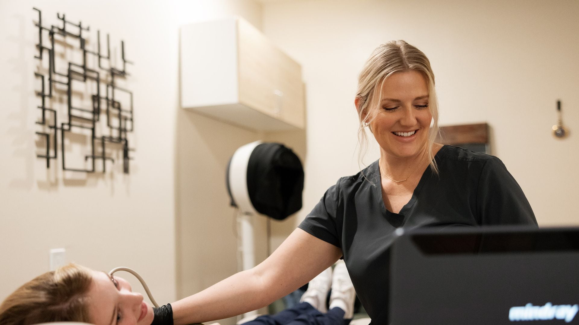
The thyroid sits just under the skin at the front of the neck, which makes it one of the organs ultrasound sees best. Using a high-frequency probe, the sonographer images the whole gland and measures each lobe. For every nodule, they record its size in three dimensions and describe five features. These five features are the entire basis of the TI-RADS score, so it is worth knowing what they mean.
| Feature | What the sonographer is describing | Why it matters |
|---|---|---|
| Composition | Is the nodule fluid (cystic), solid, or a mix? “Spongiform” means it is made up of many tiny cystic spaces, like a sponge. | Purely cystic and spongiform nodules are almost always benign. Solid nodules have more possible causes. |
| Echogenicity | How bright the solid part is compared with normal thyroid tissue: hyperechoic (brighter), isoechoic (same), hypoechoic (darker), very hypoechoic (darker than the neck muscles). | Darker nodules score higher. Hypoechoic is common and mostly benign; “very hypoechoic” carries more weight. |
| Shape | Is the nodule wider than it is tall, or taller than it is wide (measured on a cross-sectional view)? | Taller-than-wide is one of the strongest single predictors of concern, because benign nodules tend to grow along the tissue planes rather than across them. |
| Margins | Are the edges smooth, ill-defined, lobulated or irregular? Does the nodule appear to extend beyond the thyroid capsule? | Irregular or lobulated edges score points; extension outside the gland scores the most. |
| Echogenic foci | Bright spots inside the nodule: none, large “comet-tail” artifacts (benign, typical of colloid), coarse macrocalcifications, a rim of peripheral calcification, or tiny punctate foci. | Punctate (pinpoint) bright foci score the highest; comet-tail artifacts score zero and are reassuring. |
Along with these, the sonographer notes the nodule’s blood flow on Doppler and checks the lymph nodes in the neck, which matter for staging if anything is ever found.
How ACR TI-RADS scoring works
TI-RADS stands for Thyroid Imaging Reporting and Data System. The version used by most radiologists in the United States was published by the American College of Radiology in 2017. It assigns points for each of the five features, adds them up, and converts the total into a category from TR1 to TR5. Here is the full scoring table so you can follow your own report.
| Feature | Finding | Points |
|---|---|---|
| Composition | Cystic or almost completely cystic | 0 |
| Spongiform | 0 | |
| Mixed cystic and solid | 1 | |
| Solid or almost completely solid | 2 | |
| Echogenicity | Anechoic | 0 |
| Hyperechoic or isoechoic | 1 | |
| Hypoechoic | 2 | |
| Very hypoechoic | 3 | |
| Shape | Wider than tall | 0 |
| Taller than wide | 3 | |
| Margin | Smooth or ill-defined | 0 |
| Lobulated or irregular | 2 | |
| Extra-thyroidal extension | 3 | |
| Echogenic foci (all that apply) | None, or large comet-tail artifacts | 0 |
| Macrocalcifications | 1 | |
| Peripheral (rim) calcifications | 2 | |
| Punctate echogenic foci | 3 |
The points are summed and the total places the nodule in a category:
| Total points | Category | Meaning |
|---|---|---|
| 0 | TR1 | Benign |
| 2 | TR2 | Not suspicious |
| 3 | TR3 | Mildly suspicious |
| 4 to 6 | TR4 | Moderately suspicious |
| 7 or more | TR5 | Highly suspicious |
An example: a solid (2 points), hypoechoic (2 points), wider-than-tall (0) nodule with smooth margins (0) and no echogenic foci (0) totals 4 points, which is TR4. Notice that a very ordinary-looking solid, slightly dark nodule lands in TR4 purely on composition and echogenicity. That is why TR4 is common, and why “moderately suspicious” does not mean “probably cancer.” Most TR4 nodules are benign.
Why size decides what happens next
Here is the part that confuses people most. The TI-RADS category tells the radiologist how suspicious the nodule looks. The size then decides whether anything should be done about it. ACR’s recommendations:
| Category | Biopsy (FNA) recommended if | Follow-up ultrasound if | Otherwise |
|---|---|---|---|
| TR1 | Never | Not needed | No action |
| TR2 | Never | Not needed | No action |
| TR3 | 2.5 cm or larger | 1.5 cm or larger | No follow-up needed |
| TR4 | 1.5 cm or larger | 1.0 cm or larger | No follow-up needed |
| TR5 | 1.0 cm or larger | 0.5 cm or larger | No follow-up needed |
So a 1.4 cm TR4 nodule does not meet the biopsy threshold; it meets the follow-up threshold. A 0.8 cm TR5 nodule is followed, not biopsied. The logic is that small nodules, even suspicious-looking ones, rarely cause harm, and biopsying every one of them would mean a great many needles for very little benefit. The follow-up scan exists to catch the uncommon nodule that grows or changes.
How often is “follow-up”?
ACR suggests re-scanning TR3 nodules at 1, 3 and 5 years; TR4 at 1, 2, 3 and 5 years; and TR5 every year for up to 5 years. If a nodule is stable across that period, it is very unlikely to be clinically significant, and surveillance usually stops. “Growth” is defined fairly generously (a 20 percent increase in at least two dimensions, with a minimum of 2 mm, or a 50 percent increase in volume) so that normal measurement variation does not trigger alarm.
What a biopsy involves, and what the results mean
If a nodule meets the threshold, the next step is a fine-needle aspiration (FNA): under ultrasound guidance, a doctor passes a very thin needle into the nodule a few times to collect cells. It takes minutes, is done with local anesthetic or none, and feels like a blood draw in the neck. The cells are reported using the Bethesda system, which has six categories ranging from “benign” (the most common result) through “indeterminate” categories to “malignant.” Indeterminate results are where molecular testing of the sample increasingly helps avoid unnecessary surgery.
Keep in mind that even when thyroid cancer is found, the most common type (papillary) is typically slow-growing and highly treatable; for very small, low-risk cancers, active monitoring rather than surgery is now an accepted option in many centers. The conversation about what to do belongs with an endocrinologist or thyroid surgeon.
Things ultrasound does not tell you about your thyroid
- Whether the gland is working properly. Thyroid function is a blood test (TSH, and sometimes T4 and T3). A nodule can be present with completely normal function, and an under- or over-active thyroid can occur without any nodules.
- Whether a nodule is “hot.” A small minority of nodules overproduce thyroid hormone. If your TSH is low, your doctor may order a nuclear medicine uptake scan; hot nodules are almost never cancerous and are usually not biopsied.
- Whether a nodule is cancer. Ultrasound estimates risk; only cells from a biopsy can confirm it. See our article on what ultrasound can and cannot detect.
Who should be more careful
Some factors raise the likelihood that a nodule matters, and your doctor will weigh them alongside the TI-RADS score: radiation to the head or neck in childhood, a family history of thyroid cancer or certain inherited syndromes, a nodule that is growing quickly, new hoarseness or trouble swallowing, a firm or fixed lump, or enlarged lymph nodes on the same side. A nodule in a child or in a man is also looked at a little more closely, simply because nodules are less common in those groups.
Reading your own report: a short checklist
- Find the TI-RADS category for each nodule (reports list them individually if there are several; the one with the highest category drives the plan).
- Find the largest dimension of that nodule.
- Compare them with the thresholds above. Most readers will find their nodule sits in the “follow-up” or “no follow-up needed” box.
- Look for the words “stable” and “unchanged” if you have had a prior scan. Stability over time is strong reassurance.
- Bring the report to your doctor with the question: “Does this need a biopsy, a follow-up scan, or nothing?”
If you want help with the rest of the vocabulary (hypoechoic, heterogeneous, echotexture), our guide to reading an ultrasound report covers it.
Getting a thyroid ultrasound
Thyroid ultrasound needs no preparation, takes about half an hour, and at UltraVision Imaging does not require a referral. Every study is performed by an ARDMS-certified sonographer and read by a board-certified radiologist who reports each nodule with its TI-RADS category, and the written report is typically ready within 24 hours, for you and for your physician. It is a good fit for a first look at a lump you have noticed, or for the 1-, 2-, 3- or 5-year follow-up scans on a known nodule. Enter your ZIP to see times at our private clinic or for a home visit, or call (916) 751-1944.
Frequently asked questions
What does TI-RADS mean on a thyroid ultrasound?
TI-RADS (Thyroid Imaging Reporting and Data System) is a scoring system radiologists use to rate how suspicious a thyroid nodule looks. Points are given for the nodule’s composition, echogenicity, shape, margins and echogenic foci, and the total places it in a category from TR1 (benign) to TR5 (highly suspicious). The category, together with the nodule’s size, determines whether a biopsy or a follow-up scan is recommended.
Is a TI-RADS 4 thyroid nodule cancer?
Not usually. TR4 means “moderately suspicious,” and a very ordinary solid, slightly dark nodule can score TR4 on composition and echogenicity alone. Most TR4 nodules are benign. A biopsy is recommended only if a TR4 nodule is 1.5 cm or larger; smaller ones are followed with ultrasound.
Is a TI-RADS 3 nodule something to worry about?
TR3 is “mildly suspicious.” Biopsy is recommended only if the nodule is 2.5 cm or larger, and follow-up ultrasound only if it is 1.5 cm or larger. Smaller TR3 nodules need no follow-up.
When does a thyroid nodule need a biopsy?
Under ACR TI-RADS: TR3 nodules 2.5 cm or larger, TR4 nodules 1.5 cm or larger, and TR5 nodules 1 cm or larger. Your doctor may also consider risk factors such as childhood neck radiation, family history, rapid growth or suspicious lymph nodes.
How common are thyroid nodules?
Very common. High-resolution ultrasound finds nodules in a large share of adults, in many studies around half, and the proportion increases with age. Roughly 90 to 95 percent of nodules are benign.
How often should a thyroid nodule be rechecked?
ACR suggests follow-up ultrasound at 1, 3 and 5 years for TR3 nodules, at 1, 2, 3 and 5 years for TR4, and yearly for up to 5 years for TR5. Nodules that stay stable over that period are very unlikely to be significant.
Does a thyroid ultrasound show if my thyroid is working?
No. Ultrasound shows the structure of the gland and any nodules. Thyroid function is measured with blood tests such as TSH. The two are often done together.
Do I need a referral for a thyroid ultrasound?
Not at UltraVision Imaging. Thyroid ultrasound can be booked directly, needs no preparation, and the radiologist’s report, including TI-RADS categories for any nodules, is typically ready within 24 hours.
This article is general health information, not medical advice. It does not replace a conversation with your own physician, who knows your history and can interpret results in context. If you have symptoms that worry you, contact your doctor; if you think you are having a medical emergency, call 911.