
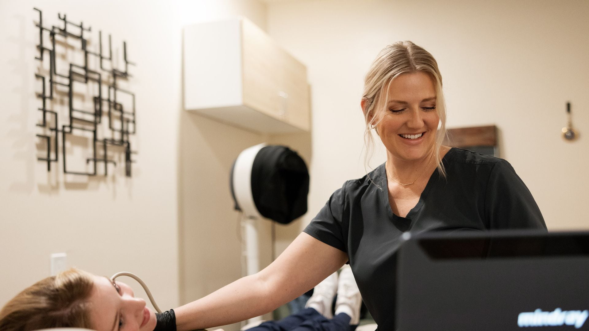
You had a thyroid ultrasound, the report described one or more nodules, and the last line said something like “recommend follow-up ultrasound in 1 year.” Now the questions start. Why wait a year? What exactly will they compare? How much growth is too much? And when does follow-up end?
If you want the basics of what a nodule is and how ACR TI-RADS scores it, start with our explainer, thyroid nodules and TI-RADS explained. This article picks up where that one ends. It focuses on follow-up: how often nodules are typically rescanned, what counts as a meaningful change, when a biopsy (FNA) is recommended, what happens after a benign biopsy, and the basics of ultrasound surveillance for people who have been treated for thyroid cancer.
Why follow-up exists at all
Most thyroid nodules are benign, and most never cause a problem. Biopsying every nodule would lead to many unnecessary procedures and inconclusive results. Surveillance is the middle path: nodules that look low-risk but are big enough to matter get rechecked over time, and a nodule that changes is reassessed. Stability over several years is itself reassuring evidence that a nodule is behaving benignly.
ACR TI-RADS follow-up intervals
The American College of Radiology’s TI-RADS system pairs a risk level (TR1 through TR5) with size thresholds. Below the follow-up size, routine rescanning is generally not recommended. Between the follow-up size and the biopsy size, rescanning is recommended. At or above the biopsy size, FNA is recommended.
| TI-RADS level | Follow-up ultrasound if | FNA recommended if | Suggested rescan schedule |
|---|---|---|---|
| TR1, benign | Not needed | Not needed | No routine follow-up |
| TR2, not suspicious | Not needed | Not needed | No routine follow-up |
| TR3, mildly suspicious | 1.5 cm or larger | 2.5 cm or larger | At 1, 3, and 5 years |
| TR4, moderately suspicious | 1.0 cm or larger | 1.5 cm or larger | At 1, 2, 3, and 5 years |
| TR5, highly suspicious | 0.5 cm or larger | 1.0 cm or larger | Every year for up to 5 years |
A few rules sit behind that table:
- Five years of stability is usually enough. The ACR TI-RADS white paper notes that imaging can generally stop at five years when a nodule has not changed, because that kind of stability indicates benign behavior.
- A higher TI-RADS level on a later scan generally prompts the next ultrasound in one year, regardless of the original schedule.
- Multiple nodules: ACR suggests reporting and following up to four of the highest-scoring nodules, and biopsying no more than the two highest-scoring nodules that meet criteria. The biggest nodule is not always the most important one.
- Your doctor may adjust. Age, a history of radiation to the head or neck, a family history of thyroid cancer, symptoms, and personal preference all reasonably change the plan.
What counts as significant growth
ACR TI-RADS defines significant enlargement as an increase of at least 20 percent and at least 2 mm in at least two dimensions, or a 50 percent or greater increase in volume. The two-millimeter floor is there because small differences in probe angle, pressure, and caliper placement can make a nodule measure a millimeter or two differently on the same day.
Here is how that plays out in two examples:
| Earlier scan | New scan | Significant growth? | Why |
|---|---|---|---|
| 12 x 10 x 9 mm | 15 x 13 x 9 mm | Yes | Two dimensions grew by at least 20 percent and at least 2 mm (12 to 15, 10 to 13); volume rose about 60 percent |
| 6 x 5 x 5 mm | 7 x 5 x 5 mm | No | Only one dimension changed, by 1 mm, which is within normal measurement variation |
| 20 x 15 x 14 mm | 22 x 16 x 14 mm | No | Changes are under 20 percent and only one dimension changed by 2 mm |
Growth alone does not mean cancer. Benign nodules often grow slowly. What growth does is trigger a fresh look: the radiologist rescores the nodule, and your doctor considers whether a biopsy, or a repeat biopsy, is warranted. Changes in features matter too, such as a new irregular margin, new tiny bright spots (punctate echogenic foci), or extension beyond the thyroid.
When FNA is recommended
Fine-needle aspiration uses a thin needle, usually guided by ultrasound, to collect cells. It is typically recommended when:
- A nodule meets or passes the FNA size threshold for its TI-RADS level
- A followed nodule grows significantly or its features become more suspicious
- There is an abnormal-looking lymph node in the neck, which may be sampled directly
- Your doctor has other reasons, such as risk factors or symptoms
Results are reported with the Bethesda system, from category I (nondiagnostic) through VI (malignant). A nondiagnostic result usually means the sample did not have enough cells and the biopsy is repeated. An indeterminate result (category III or IV) may lead to a repeat FNA, molecular testing of the sample, or surgery, depending on the situation. We do not perform biopsies; your doctor arranges FNA with an endocrinologist, radiologist, or surgeon who does them.
After a benign biopsy
A benign (Bethesda II) result is reassuring. False-negative results are uncommon but do happen; published studies report rates in roughly the low single digits, with ranges up to about 10 percent in some series. That is why follow-up after a benign biopsy depends on how the nodule looked on ultrasound. The 2015 American Thyroid Association guidelines describe this approach:
| Ultrasound appearance | Typical follow-up after a benign FNA (2015 ATA) |
|---|---|
| High suspicion pattern | Repeat ultrasound and repeat FNA within 12 months |
| Low to intermediate suspicion | Repeat ultrasound in 12 to 24 months; if it grows significantly or develops new suspicious features, consider repeat FNA or continued observation |
| Very low suspicion (such as spongiform) | Surveillance of limited value; if repeated, no sooner than 24 months |
| Two benign FNAs of the same nodule | Ultrasound surveillance for cancer risk is generally no longer needed |
Even when cancer surveillance ends, some people still get an occasional ultrasound because a large nodule is causing pressure symptoms or cosmetic concerns, or because new nodules may appear elsewhere in the gland.
Neck lymph nodes: part of every good follow-up
A thyroid ultrasound should include a look at the lymph nodes in the neck, because papillary thyroid cancer most often spreads there first. Normal nodes are usually oval with a bright fatty center (the hilum). Features that raise concern include:
- A round rather than oval shape
- Loss of the fatty hilum
- Tiny bright calcifications
- Fluid-filled (cystic) areas
- Blood flow around the edge instead of through the center
- A node that looks brighter than usual, resembling thyroid tissue
Lymph nodes swell for ordinary reasons too, like a cold or sore throat, so a single slightly enlarged node is often reactive. Persistent or suspicious nodes may be sampled with FNA, sometimes with a thyroglobulin measurement on the needle wash.
Surveillance after thyroid cancer treatment
For people treated for differentiated thyroid cancer (papillary, follicular, or oncocytic), neck ultrasound is a core part of long-term follow-up. It checks the thyroid bed, where the gland used to be, and the lymph node regions of the neck. The American Thyroid Association published updated guidelines for differentiated thyroid cancer in 2025, building on the 2015 version. In broad strokes:
- A first neck ultrasound is commonly done within the first year after surgery, often around 6 to 12 months.
- After that, the frequency depends on the original risk level and how well treatment worked, often judged with blood tests for thyroglobulin and thyroglobulin antibodies. People with an excellent response may need ultrasound less and less often over time.
- If only one lobe was removed, ultrasound also follows the remaining lobe for new nodules.
- For some small, low-risk papillary cancers, specialists may offer active surveillance with regular ultrasound instead of immediate surgery.
Your endocrinologist or surgeon sets this schedule; it is individual. Blood tests like thyroglobulin and TSH are drawn at a lab such as Quest or Labcorp, not at an ultrasound visit, since we do not draw blood.
Making follow-up scans comparable
Follow-up only works if the new scan can be compared fairly with the old one. A few practical steps help:
- Bring or send your prior report, and images if you have them. The radiologist compares measurements and features; without prior studies, growth cannot be judged.
- Keep a simple record: the date of each scan, the nodule location (for example, right lobe, lower pole), its TI-RADS level, and three measurements.
- Keep to the recommended interval. Scanning too soon adds noise; skipping years makes changes harder to interpret.
- Tell us which nodule your doctor is following, especially if you have several.
- Remember that ultrasound does not test thyroid function. If you have symptoms of an overactive or underactive thyroid, ask your doctor about blood tests.
When to seek urgent care
Thyroid nodules rarely cause emergencies, but go to an ER or call 911 for:
- Neck swelling that grows quickly, with trouble breathing, noisy breathing, or difficulty swallowing
- Rapidly expanding swelling or bleeding after a biopsy
- Severe neck pain with fever and feeling very unwell
Contact your doctor promptly, rather than waiting for the next scheduled scan, for a new lump you can feel, persistent hoarseness, or a nodule that seems to be growing between scans.
Getting your follow-up scan with us
A thyroid ultrasound can be booked directly; no referral is needed, though bringing your doctor’s referral and prior reports helps the radiologist answer the right question. There is no prep: wear an open neckline, and avoid turtlenecks or high collars. You can book online at our clinic, or we bring the scan to you in our California service areas. It is a flat rate paid when you book, with no facility fee, and HSA/FSA is accepted.
A board-certified radiologist reads every study, and the report is ready within 24 hours. It goes to you and, if named, your doctor. Our guide to ultrasound reports can help with terminology, and your doctor will help you decide what the findings mean for your next step.
Getting a scan with us: our thyroid ultrasound page explains what the scan covers and how to prepare. You can book online for our clinic, or we bring the scan to you at home. A board-certified radiologist reads every study, and the report is ready within 24 hours. Your own doctor interprets the results for you.
Sources
- Radiopaedia: ACR Thyroid Imaging Reporting and Data System (ACR TI-RADS)
- Haugen BR et al. 2015 American Thyroid Association Management Guidelines for Adult Patients with Thyroid Nodules and Differentiated Thyroid Cancer (Thyroid, via PMC)
- American Thyroid Association: New ATA Management Guidelines for Adult Patients with Differentiated Thyroid Cancer (2025)
- American Thyroid Association, Clinical Thyroidology for the Public: 2025 differentiated thyroid cancer guidelines summary
- American Thyroid Association, Clinical Thyroidology for the Public: Follow-up of nodules with benign biopsy results
Frequently asked questions
How often should a thyroid nodule be rescanned?
Under ACR TI-RADS, TR3 nodules of 1.5 cm or larger are usually rescanned at 1, 3, and 5 years; TR4 nodules of 1.0 cm or larger at 1, 2, 3, and 5 years; and TR5 nodules of 0.5 cm or larger every year for up to 5 years. TR1 and TR2 nodules generally need no routine follow-up. Your doctor may adjust the schedule.
How much growth in a thyroid nodule is significant?
ACR TI-RADS defines significant growth as an increase of at least 20 percent and at least 2 mm in at least two dimensions, or a volume increase of 50 percent or more. Smaller changes are often measurement variation. Growth prompts reassessment but does not by itself mean cancer.
When can thyroid nodule follow-up stop?
The ACR TI-RADS white paper notes that imaging can generally stop after five years if the nodule has not changed. After two benign biopsies of the same nodule, 2015 ATA guidance says ultrasound surveillance for cancer risk is generally no longer needed. Your doctor makes the final call.
Do I still need ultrasounds after a benign thyroid biopsy?
Often, yes, depending on how the nodule looks. The 2015 ATA guidelines suggest repeat ultrasound and FNA within 12 months for a high suspicion pattern, repeat ultrasound in 12 to 24 months for low to intermediate suspicion, and little or infrequent surveillance for very low suspicion nodules.
How are thyroid cancer survivors monitored with ultrasound?
Neck ultrasound of the thyroid bed and lymph nodes is a core part of follow-up, commonly first within 6 to 12 months after surgery and then at intervals based on risk and response to treatment, alongside thyroglobulin blood tests. Your endocrinologist sets the schedule.
Do I need to prepare for a thyroid ultrasound?
No prep is needed. Wear an open neckline. You can book directly without a referral, and a board-certified radiologist reads every study with the report ready within 24 hours.
This article is general health information, not medical advice. It does not replace a conversation with your own physician, who knows your history and can interpret results in context. If you have symptoms that worry you, contact your doctor; if you think you are having a medical emergency, call 911.