
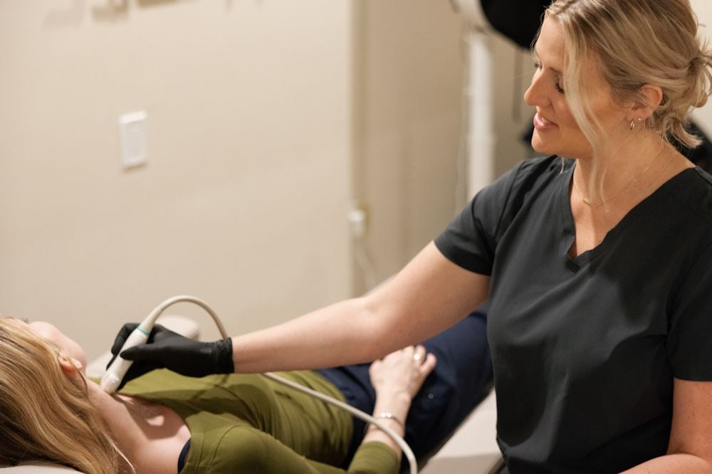
A swollen, tender calf is one of the most common reasons a concierge or house-call physician reaches for imaging. The patient is usually at home, often older, sometimes recently operated on or just off a long flight, and almost always reluctant to spend six hours in an emergency department for a test. The physician’s job is to decide quickly and safely: is this a patient who can have a diagnostic venous duplex where they are, or one who needs the emergency department now?
This guide summarizes how guideline-based pathways approach suspected deep vein thrombosis, where a mobile venous duplex fits, and the red flags that should send a patient to the hospital regardless of convenience. It is general information for clinicians, not a substitute for your own judgment about the patient in front of you.
Start with the red flags, not the duplex
Before deciding where to image, decide whether the patient needs an emergency evaluation for something other than the leg. Features that point to the emergency department include:
- Shortness of breath, chest pain, a new rapid heart rate, low oxygen saturation, fainting or coughing blood, which raise concern for pulmonary embolism.
- A cold, pale or pulseless limb, or severe pain out of proportion to the exam, which raise concern for arterial compromise or compartment syndrome.
- Massive swelling with a bluish or mottled limb (phlegmasia).
- Signs of systemic infection, such as fever with spreading redness, that may point to cellulitis or an abscess needing urgent care.
- An unstable patient, or one who cannot be reliably followed up within the next day.
None of these patients should wait for a scheduled or mobile study. A mobile duplex is designed for the stable patient with an isolated limb question.
Estimate pretest probability
Most pathways begin with a structured estimate of pretest probability. The Wells score for DVT is the most widely used. It assigns points for active cancer, paralysis or recent immobilization of the leg, recent bed rest or major surgery, localized tenderness along the deep veins, entire-leg swelling, calf swelling more than 3 cm compared with the other side, pitting edema in the symptomatic leg, collateral superficial veins and a previous DVT, and it subtracts points when an alternative diagnosis is at least as likely.
The American Society of Hematology’s 2018 guideline on VTE diagnosis built its recommendations around that estimate. In broad terms:
- Low probability: a highly sensitive D-dimer is often the first test. A negative result can exclude DVT without imaging in many patients.
- Intermediate probability: either a D-dimer first or ultrasound first, depending on the setting.
- High probability: proceed to ultrasound. A D-dimer alone is not enough to exclude DVT.
For concierge practices, the D-dimer step is often the practical bottleneck. Some practices draw it in the home and send it to a reference lab; others go straight to ultrasound because a same-day lab result is not realistic. Either is a reasonable path when it follows a defined protocol.
Proximal versus whole-leg ultrasound
Two ultrasound strategies are described in the literature. A proximal compression study evaluates the femoral and popliteal veins; if negative in a patient who is not low risk, it is often repeated in about a week to catch calf clots that propagate. A whole-leg (complete) duplex evaluates the proximal and calf veins in one session, which avoids the repeat visit but finds more isolated distal clots whose management varies.
For a house-call population, a complete duplex at the first visit is often the most practical. It avoids a second home visit and gives the physician a full picture. Ask your imaging provider which protocol it performs and what the report will include.
Where a mobile venous duplex fits
A mobile duplex is a good fit when the patient is stable, the question is limited to the limb, and the physician wants a formal study without moving the patient. Typical examples:
- An older patient with unilateral calf swelling and no respiratory symptoms.
- A post-operative patient recovering at home or in an aftercare residence, cleared by their surgeon for outpatient evaluation.
- A traveling member with leg swelling after a long flight, staying at a hotel.
- A patient with known varicose veins and a new tender cord, where the question is superficial thrombophlebitis and its distance from the deep system.
Timing: a fast visit is not a fast report
Physicians should be clear about what “STAT” means when ordering mobile imaging. A provider may be able to reach the patient the same day, but the radiologist’s written report follows its own timeline, often within 24 hours. If treatment decisions cannot wait for the written report, discuss with the provider before the visit how urgent findings will be communicated, and to whom. If no rapid communication path exists, a high-probability patient may be better served in the emergency department, where imaging and treatment happen in the same building.
Some practices begin empiric anticoagulation while awaiting imaging in selected patients with high pretest probability and low bleeding risk, consistent with guideline language. That is a clinical decision for the treating physician.
What to include in the order
A good order speeds the study and sharpens the report:
- Which limb, or both, and whether upper or lower extremity.
- The clinical question: suspected DVT, follow-up of known DVT, superficial thrombophlebitis, or swelling of unclear cause.
- Relevant history: recent surgery or travel, cancer, prior DVT, current anticoagulation.
- Who should receive urgent findings, with a direct number that will be answered.
After the report
The radiologist’s report will describe each venous segment evaluated, the compressibility and flow, and any thrombus with its location and extent. It may also note incidental findings, such as a Baker’s cyst, a hematoma or enlarged lymph nodes, that explain the symptoms. The ordering physician interprets those findings in context and directs treatment and follow-up.
Special populations
Pregnant and postpartum patients
Pregnancy and the weeks after delivery carry an increased risk of venous thromboembolism, and leg swelling is common in pregnancy for other reasons too. Duplex ultrasound is safe in pregnancy. Pregnancy-specific pathways exist, and left-sided symptoms and iliac vein involvement are more common, which can affect the choice of protocol. These patients are usually managed with the obstetric team.
Patients with cancer
Active cancer is one of the strongest risk factors for VTE and adds points to the Wells score. Concierge oncology patients receiving treatment at home may have both leg and arm symptoms, particularly with central lines. A low threshold for duplex is reasonable, and emergency evaluation is appropriate for any respiratory symptoms.
Follow-up after a negative study
A negative complete duplex in a patient with persistent or worsening symptoms does not end the conversation. Guidelines describe repeat imaging in selected patients, particularly after a negative proximal-only study or when symptoms progress. The ordering physician decides whether and when to repeat, and a mobile repeat study can be scheduled at the same location.
Ordering a mobile venous duplex
UltraVision Imaging performs lower and upper extremity venous duplex studies at patients’ homes, hotels and offices for concierge, house-call and private-medicine practices across Los Angeles, San Diego County, Orange County and Palm Springs, the Bay Area and the Sacramento region. A board-certified radiologist reads every study, and the report goes to the ordering physician. For the fastest timing, call the local line for your region on our page for physicians. Our team will be in touch to coordinate scheduling.
Frequently asked questions
Can a DVT ultrasound be done at the patient’s home?
Yes. A complete venous duplex can be performed with a portable diagnostic system at a home, hotel or office, and read by a radiologist.
When should a patient with leg swelling go to the emergency department instead?
When there are signs of pulmonary embolism, limb-threatening ischemia, phlegmasia, systemic infection, instability, or no reliable follow-up. Those patients should not wait for mobile imaging.
Is a D-dimer needed before ultrasound?
Guidelines use D-dimer mainly in patients with low or intermediate pretest probability. In high-probability patients, ultrasound is the next step.
What is the difference between a proximal and a whole-leg study?
A proximal study evaluates the femoral and popliteal veins and may need a repeat in about a week. A whole-leg study adds the calf veins in one session.
How fast is the report?
The written radiologist report is usually available within 24 hours. A same-day visit does not mean a same-day report; agree on urgent-finding communication in advance.
Can upper extremity veins be scanned at home?
Yes. Upper extremity venous duplex, for example in a patient with a PICC line or arm swelling, can be performed with a portable system.
Sources
- Lim W et al. American Society of Hematology 2018 guidelines for management of venous thromboembolism: diagnosis of VTE. Blood Adv 2018
- Wells PS et al. Evaluation of D-dimer in the diagnosis of suspected deep-vein thrombosis. N Engl J Med 2003
- ACR Appropriateness Criteria: Suspected Lower Extremity Deep Vein Thrombosis
- RadiologyInfo.org (ACR and RSNA): Venous ultrasound
- CDC: Venous thromboembolism (blood clots)
This article is general information for licensed clinicians and practice staff. It is not medical or legal advice and does not replace clinical judgment. UltraVision Imaging performs diagnostic ultrasound; a board-certified radiologist interprets every diagnostic study, and the ordering physician directs care.