
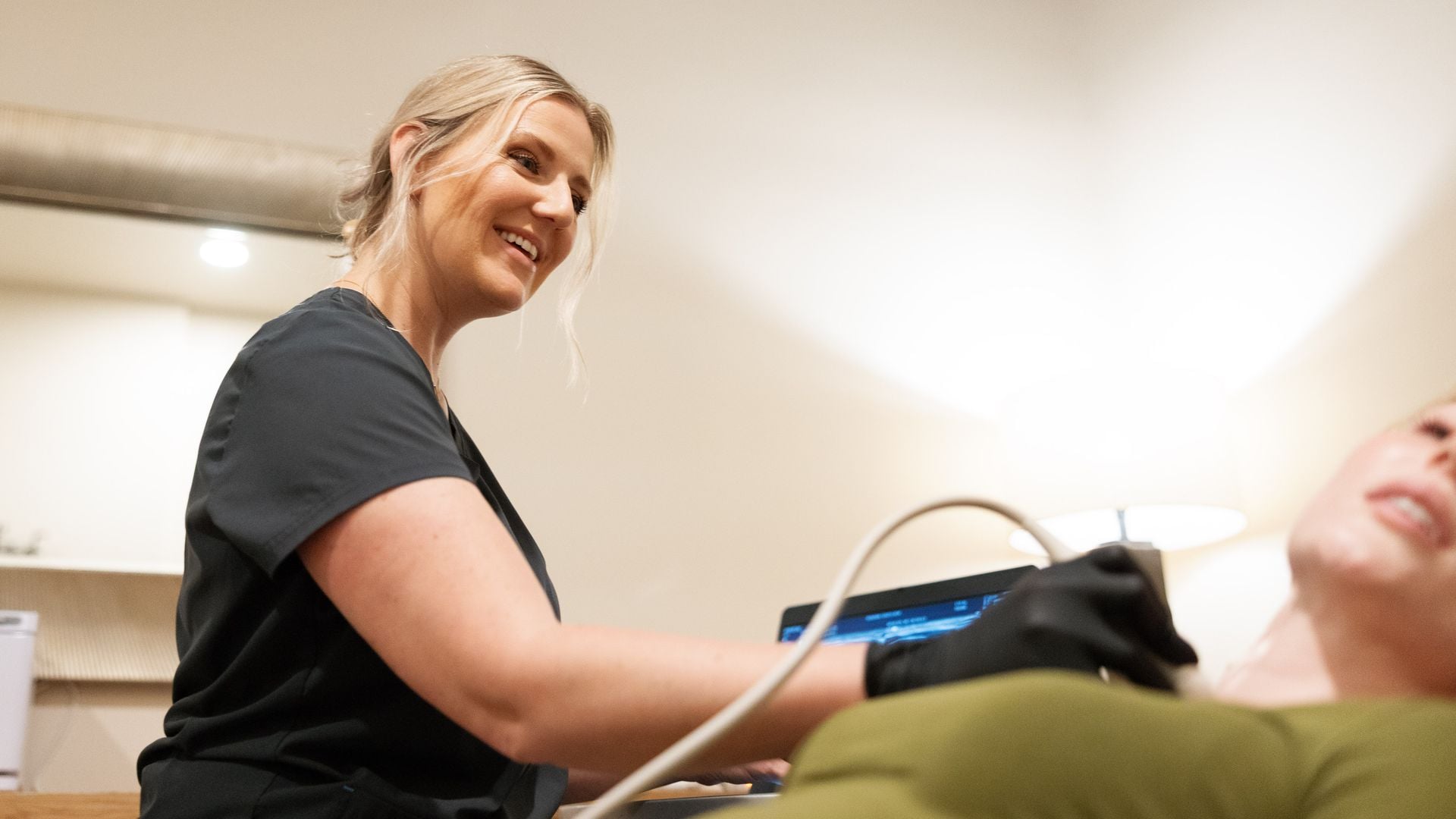
Point-of-care ultrasound has changed bedside medicine. A physician with a handheld probe can answer a focused question in minutes, during the visit, without sending the patient anywhere. That speed is real and valuable. It also creates a recurring question for concierge and house-call physicians: when is my own bedside scan enough, and when does the patient need a complete diagnostic study with a radiologist’s report?
The two are not competing versions of the same test. They are different exams built for different jobs. Knowing where the line falls protects the patient, keeps the record clean for follow-up, and helps you avoid owning an interpretation you never intended to make.
What POCUS is designed to do
Point-of-care ultrasound is performed and interpreted by the treating clinician at the bedside to answer a specific, usually binary question that changes immediate management. Professional guidelines describe it this way: a focused exam integrated into the physical examination, not a comprehensive survey of an organ system.
Typical concierge and house-call uses include:
- Is the bladder distended, and roughly how much is in it?
- Is there a pericardial effusion?
- Are there B-lines suggesting interstitial fluid in a short-of-breath patient?
- Is there free fluid in the abdomen?
- Guidance for a joint aspiration or a peripheral IV.
In each case, the physician is looking for the presence or absence of a finding to decide what to do next. POCUS is excellent at that, and in skilled hands it can speed decisions dramatically.
What a formal diagnostic ultrasound adds
A formal diagnostic study is a complete examination performed to a published protocol, usually by a registered sonographer, with a full set of archived images and a written report from a radiologist. The protocol matters. A complete abdominal ultrasound documents the liver, gallbladder, bile ducts, pancreas, spleen, kidneys and aorta with standard views and measurements. A complete venous duplex for suspected DVT evaluates the deep veins along a defined length of the limb with compression and Doppler.
That completeness is what makes the study useful beyond the moment:
- Comparability. A standardized study can be compared with the next one. A thyroid nodule measured to protocol today can be followed with confidence in a year.
- A second set of eyes. The radiologist reads every image without having been in the room, which catches findings outside the question that prompted the order.
- A defensible record. The written report documents what was seen, what was not, and what the radiologist recommends, such as follow-up imaging.
- Structured risk language. Many reports use standardized systems, such as ACR TI-RADS for thyroid nodules or O-RADS for adnexal findings, that tie a finding to an evidence-based next step.
A practical way to decide
A simple test helps: will anyone need to rely on this exam after today? If the scan answers an immediate question and is unlikely to be referenced again, a bedside exam may be enough. If the result will drive a referral, a treatment decision, a follow-up interval or a comparison months from now, order the formal study.
Some common situations, framed the way concierge physicians tend to meet them:
Suspected deep vein thrombosis
Limited compression techniques at the bedside are well described in emergency medicine, but a negative focused exam does not carry the same weight as a complete duplex, particularly for calf veins and when pretest probability is not low. Guideline-based pathways for suspected DVT generally combine clinical probability, D-dimer and a diagnostic ultrasound. When the question is whether to treat, most physicians order the full venous study.
A palpable neck lump or thyroid nodule
A bedside look can confirm that a lump is thyroid rather than a lymph node. Deciding whether a nodule needs fine-needle aspiration or follow-up depends on composition, echogenicity, shape, margins, echogenic foci and size, which is exactly what a TI-RADS report documents. That is a formal study.
Right upper quadrant pain
POCUS can show gallstones quickly. A complete abdominal study also evaluates the bile ducts, liver and pancreas, which matters when the presentation is not classic or labs are abnormal.
Wellness and longevity screening
Screening exams, such as an abdominal aorta measurement in an eligible patient, should be complete and documented, because they become the baseline for every future comparison.
The liability question
When a physician performs and interprets an ultrasound, that physician owns the interpretation. Incidental findings outside the question you were asking still belong to you if they were visible on the images you obtained. That is not a reason to avoid POCUS. It is a reason to be deliberate about scope, to document focused exams as focused, and to order a formal study when the clinical question is broad.
Many practices find a hybrid works best: the physician uses POCUS to triage and decide urgency, then orders the formal study for anything that needs a durable answer.
Building a hybrid workflow in your practice
Practices that use both POCUS and formal studies well usually follow a simple pattern. The physician performs a focused bedside exam when it will change what happens in the next hour: whether to send a patient to the emergency department, whether a bladder needs catheterizing, whether a joint can be aspirated. The physician documents it as a focused exam, states the question and the answer, and orders a formal study whenever the result needs to be relied on later. Over time, this pattern becomes second nature and keeps the scope of each exam clear.
Training and quality assurance for physician-performed POCUS
Physicians who use POCUS regularly benefit from a structured approach to training and quality review. Specialty societies describe pathways that include didactic learning, supervised scans and ongoing review of saved images. Saving representative images, even for focused exams, allows later review and supports the documentation. Some practices periodically compare their bedside impressions with the formal reports on the same patients, which is a practical way to check calibration.
Explaining the difference to patients
Patients who have had a bedside scan sometimes ask why they need “another ultrasound.” A simple explanation helps: the quick scan in the office answered one question right away, and the full study looks at the whole area in a standard way, so a radiologist can read it and it can be compared with future scans. Most patients appreciate knowing that the second study adds something specific rather than repeating the first.
Equipment differences that matter
Handheld devices have improved dramatically, but diagnostic-grade portable systems still offer advantages for complete studies: multiple probe types for deep and superficial structures, more sensitive color and spectral Doppler for vascular work, finer measurement tools, and full image archiving for comparison. Those capabilities are what allow a mobile formal study to match the protocol a hospital department follows.
Getting the formal study without sending the patient out
The old trade-off was convenience versus completeness. A formal study meant sending a concierge patient into a hospital imaging department. Mobile diagnostic ultrasound removes that trade-off. An ARDMS-registered sonographer performs the complete protocol at the patient’s home, hotel or office, a board-certified radiologist reads it, and the report goes to the ordering physician, usually within 24 hours.
UltraVision Imaging performs formal diagnostic studies for concierge, house-call and private-medicine practices in Los Angeles, San Diego County, Orange County and Palm Springs, the Bay Area and the Sacramento region. See all regions on our page for physicians. Our team will be in touch to coordinate scheduling.
Frequently asked questions
Is POCUS less accurate than a formal ultrasound?
Not for the focused question it is designed to answer in trained hands. It is narrower in scope: it does not survey the whole organ system to a protocol and does not produce a radiologist report.
Can a formal study be done at the patient’s home?
Yes. Portable diagnostic systems allow complete protocols, such as abdominal, venous duplex, thyroid and pelvic studies, to be performed at a home, hotel or office.
Who interprets a mobile diagnostic study?
A board-certified radiologist reviews the images and issues a written report to the ordering physician. The sonographer does not interpret.
Should I document a POCUS exam as limited?
Many physicians document focused exams as focused or limited and state the question answered. Follow your institution’s or carrier’s guidance.
What is TI-RADS?
The ACR Thyroid Imaging Reporting and Data System, a scoring system that links ultrasound features of a thyroid nodule to recommendations for biopsy or follow-up.
Does a negative bedside compression exam rule out DVT?
Not in every case. Guideline pathways combine clinical probability, D-dimer and diagnostic ultrasound, and a complete duplex evaluates more of the venous system than a focused exam.
Sources
- ACEP: Ultrasound Guidelines: Emergency, Point-of-Care, and Clinical Ultrasound Guidelines in Medicine
- Tessler FN et al. ACR Thyroid Imaging, Reporting and Data System (TI-RADS). J Am Coll Radiol 2017
- Lim W et al. American Society of Hematology 2018 guidelines for VTE: diagnosis of venous thromboembolism. Blood Adv 2018
- AIUM: Practice parameters for ultrasound examinations
- RadiologyInfo.org (ACR and RSNA): General ultrasound
This article is general information for licensed clinicians and practice staff. It is not medical or legal advice and does not replace clinical judgment. UltraVision Imaging performs diagnostic ultrasound; a board-certified radiologist interprets every diagnostic study, and the ordering physician directs care.