
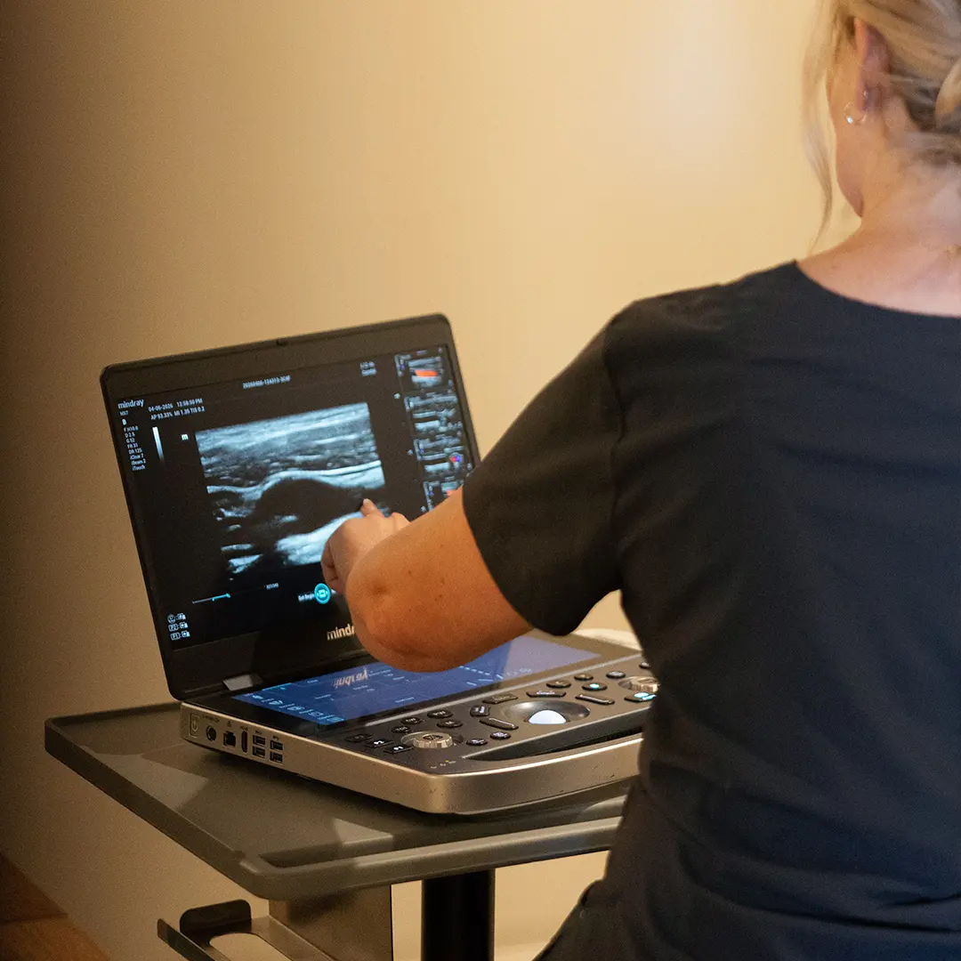
Since September 2024, every mammogram report sent to a patient in the United States has to say whether the breast tissue is dense or not dense. For many people, that letter is the first time they have heard the term, and it raises an obvious question: if dense tissue can hide cancer on a mammogram, should I get an ultrasound instead? This article explains what breast density is, what the notice means in general, what the major guidelines say about screening, and where breast ultrasound fits.
Let us be clear about the most important point first. Breast ultrasound does not replace a mammogram. Mammography is the screening test with the strongest evidence behind it, and ultrasound, when it is used, is an addition to mammography, not a substitute. Decisions about which tests you need, and when, belong to you and your own doctor, who can weigh your history, family history and personal risk.
What breast density is
Breasts are made of fatty tissue and fibroglandular tissue (milk glands, ducts and supportive tissue). Density describes how much of the breast is fibroglandular compared with fat, as it appears on a mammogram. It is not something you can feel, and it is not related to breast size or firmness. Density tends to decrease with age and can change with hormones, pregnancy and weight.
Density matters for two reasons, according to the FDA. Dense tissue and many cancers both look white on a mammogram, so dense tissue can make cancers harder to see. And dense tissue is itself associated with a higher risk of developing breast cancer. Dense breasts are common: the FDA notes that roughly half of U.S. women over 40 have dense breasts on mammography, and the U.S. Preventive Services Task Force (USPSTF) says nearly half of all women do.
The four density categories
Radiologists sort breast density into four categories, often shown as letters A through D. The descriptions below follow the wording the FDA lists for reports to providers.
| Category | Description on the report | Classed as |
|---|---|---|
| A | The breasts are almost entirely fatty | Not dense |
| B | There are scattered areas of fibroglandular density | Not dense |
| C | The breasts are heterogeneously dense, which may obscure small masses | Dense |
| D | The breasts are extremely dense, which lowers the sensitivity of mammography | Dense |
What category you fall into is a description of how the tissue looks on that mammogram. It is not a diagnosis, and what it means for your screening plan is a conversation for your doctor, who can consider density together with your age, family history, genetics and prior biopsies.
The FDA breast density notification
The FDA’s update to the Mammography Quality Standards Act (MQSA) regulations took effect on September 10, 2024. It requires mammography facilities to include a breast density assessment in the report to the provider and a plain-language statement in the summary letter to the patient. For people whose tissue is dense, the letter explains, in short, that dense tissue makes cancer harder to find on a mammogram and raises breast cancer risk; that in some people with dense tissue, other imaging tests in addition to a mammogram may help find cancers; and that you should talk to your healthcare provider about breast density, your risk and your individual situation.
Notice what the letter does and does not say. It says other tests may help some people. It does not say everyone with dense breasts needs one, and it does not say which test. It directs you to your provider. That is the right next step: bring the letter to your doctor and ask what it means for you.
What the USPSTF recommends
The USPSTF updated its breast cancer screening recommendation on April 30, 2024. In plain terms:
| Group | USPSTF recommendation | Grade |
|---|---|---|
| Women 40 to 74 at average risk | Screening mammography every 2 years | B (recommended) |
| Women 75 and older | Evidence is insufficient to weigh benefits and harms of screening mammography | I statement |
| Women with dense breasts and an otherwise negative mammogram | Evidence is insufficient to weigh benefits and harms of supplemental screening with breast ultrasound or MRI | I statement |
An “I statement” is not a recommendation for or against. It means the Task Force found too little evidence on health outcomes to decide. The USPSTF noted that no studies of supplemental screening reported outcomes across multiple screening rounds, and it advises women with dense breasts to talk with their clinicians about options. The recommendation does not apply to people at high risk, such as those with a BRCA1 or BRCA2 variant, a history of high-dose chest radiation at a young age, or a prior breast cancer or high-risk lesion. Their screening plans are set differently, with their doctors.
Other groups have taken different positions. The American College of Radiology (ACR), for example, published recommendations in 2023 stating that for women with dense breasts who want supplemental screening, breast MRI is recommended, and that contrast-enhanced mammography or ultrasound could be considered for those who qualify for but cannot have MRI. Guidelines differ, which is one more reason the decision should be made with a physician who knows you.
Where breast ultrasound fits
Breast ultrasound is used in two broad ways, and it helps to keep them separate.
Diagnostic (targeted) ultrasound
This is the most common use. A person or their doctor notices something specific: a lump, an area of thickening, focal pain, nipple discharge, or a finding on a mammogram that needs a closer look. Ultrasound is very good at telling whether a lump is filled with fluid (a cyst) or is solid, and at showing its shape and edges. According to the ACR Appropriateness Criteria for palpable breast masses (2022 update), ultrasound is usually the first test for a lump in women under 30; for women 30 to 39, ultrasound, diagnostic mammography and tomosynthesis are all usually appropriate; and for women 40 and older, diagnostic mammography or tomosynthesis is generally the starting point, with ultrasound often added. Our post on breast lumps and BI-RADS on ultrasound explains how radiologists report these studies. What a finding means for you is for your doctor to explain.
Supplemental screening ultrasound
This means scanning the whole breast in someone without symptoms, in addition to a mammogram, usually because of dense tissue. Research shows it can find some additional cancers, and also that it produces more callbacks and biopsies for findings that turn out not to be cancer. In the ACRIN 6666 trial (Berg and colleagues, JAMA 2012), which enrolled women with dense breasts and at least one other risk factor, adding ultrasound to mammography found about 4.3 additional cancers per 1,000 women screened per year, while about 5 percent of women needed a biopsy because of ultrasound findings. That trade-off, more cancers found alongside more false alarms, is exactly what you and your doctor should weigh.
| Mammogram | Breast ultrasound | |
|---|---|---|
| Role in screening | The standard screening test with the strongest evidence | Supplemental only, by physician decision; does not replace a mammogram |
| Radiation | Low-dose X-ray | None |
| Strengths | Detects calcifications, which can be an early sign of some cancers; standardized screening program | Distinguishes cysts from solid lumps; useful in dense tissue and in younger patients |
| Limitations | Less sensitive in dense tissue | Can miss calcifications; more false positives when used for screening; results depend on operator skill |
| Typical use | Routine screening; diagnostic workup | Evaluating a lump or a mammogram finding; supplemental screening for some people |
Ultrasound and mammography see different things. Mammograms are good at showing tiny calcifications; ultrasound often cannot. That is the core reason ultrasound cannot stand in for a mammogram, no matter how dense the tissue is.
Questions to bring to your doctor
If your mammogram letter says your tissue is dense, or if you are considering a breast ultrasound, these questions can help structure the conversation:
- “What density category did my mammogram show, and how does that fit with my overall risk?”
- “Based on my age, family history and any genetic results, am I considered average or higher risk?”
- “Would you recommend any additional imaging for me, and if so, which test and why?”
- “If I have an ultrasound, what are the chances it finds something that needs more tests?”
- “Should I keep my regular mammogram schedule no matter what else I do?” (Ultrasound does not replace it.)
- “Is there anything in my history, such as a prior biopsy, that changes how I should be screened?”
Our earlier post on breast ultrasound and early detection covers more background. Your physician is the one who puts it together for you.
Signs to report promptly, and when it is urgent
Whatever your density, do not wait for a scheduled screening if you notice a change. Contact your doctor promptly about:
- A new lump or thickening in the breast or armpit
- Skin dimpling, puckering, or an orange-peel texture
- A nipple that turns inward when it did not before, or a rash or scaling on the nipple
- Nipple discharge, especially if it is bloody or happens without squeezing
- A change in breast size or shape, or redness and swelling over part of the breast
Redness, warmth and swelling can come from infection, but they can also be a sign of other conditions, so they always deserve a doctor’s evaluation. Seek care the same day, through your doctor or urgent care, if a breast is red, hot and painful with fever, especially if you are breastfeeding. Call 911 or go to the emergency department if you feel very unwell with high fever, confusion, rapid heartbeat or trouble breathing. We do not provide emergency or after-hours reads, and a scheduled ultrasound is not the right path for symptoms like these.
What our breast ultrasound is, and is not
When a breast ultrasound is right for you, here is how it works with us. Our sonographer performs the scan; a board-certified radiologist reads every study and writes the report, which is ready within 24 hours; and your own doctor interprets that report for you in the context of your history, exam and any mammograms. We never diagnose, and the sonographer cannot tell you what they see during the visit. Our breast ultrasound is not a screening mammogram and does not replace one. If you are due for a mammogram, keep that appointment.
Prep is simple: on the day of your scan, do not apply lotion, powder or deodorant to the chest or underarms. Wear a two-piece outfit so you only need to undress from the waist up. If you’ve had earlier breast imaging, keep those reports handy for your doctor, who can compare them with the new one. Our post on how to prepare for an ultrasound covers prep for every scan.
Getting your scan with us
You can book a breast ultrasound directly online; no referral is needed. The booking form asks for the name of the doctor who should receive the report (type “None” if you do not have one) and offers an optional referral upload if your doctor gave you one. Payment is a flat rate paid when you book, with no facility fee, and HSA/FSA cards are accepted. We see patients at our clinic and also bring the scan to you at home in our California service areas. When you are ready, book your appointment here. We strongly suggest talking with your doctor first about whether ultrasound is the right test for your situation.
Getting a scan with us: our breast ultrasound page explains what the scan covers and how to prepare. You can book online for our clinic, or we bring the scan to you at home. A board-certified radiologist reads every study, and the report is ready within 24 hours. Your own doctor interprets the results for you.
Sources
- U.S. FDA: Understanding Breast Density
- U.S. FDA: Final Rule to Amend the Mammography Quality Standards Act (MQSA)
- U.S. FDA: FDA updates mammography regulations to require reporting of breast density information
- USPSTF: Breast Cancer Screening, final recommendation (April 30, 2024)
- ACR Appropriateness Criteria: Palpable Breast Masses, 2022 Update (J Am Coll Radiol, 2023)
- Monticciolo DL, et al. Breast Cancer Screening for Women at Higher-Than-Average Risk: Updated ACR Recommendations (J Am Coll Radiol, 2023)
- Summary of Berg WA, et al. ACRIN 6666 (JAMA, 2012): supplemental ultrasound and MRI in dense breasts
- RadiologyInfo (ACR/RSNA): How to Read Your Mammography Report
Frequently asked questions
Can a breast ultrasound replace my mammogram?
No. Breast ultrasound does not replace a mammogram. Mammography is the screening test with the strongest evidence, and it shows calcifications that ultrasound often cannot. When ultrasound is used for screening, it is an addition to mammography, decided with your doctor.
What does it mean if my mammogram letter says my breasts are dense?
It means the mammogram showed a high proportion of fibroglandular tissue, which is very common. Dense tissue can make cancer harder to see on a mammogram and is associated with higher risk. The letter recommends talking with your healthcare provider about your density, your risk and your individual situation.
Does the USPSTF recommend ultrasound for dense breasts?
The USPSTF 2024 recommendation found the evidence insufficient to weigh the benefits and harms of supplemental ultrasound or MRI for women with dense breasts and an otherwise negative mammogram. That is neither a recommendation for nor against. It recommends mammography every 2 years for women 40 to 74.
What can a breast ultrasound show?
Ultrasound is good at showing whether a lump is fluid-filled or solid and at describing its shape and edges. It is commonly used to look at a lump or a mammogram finding. A board-certified radiologist reads the images and writes the report, and your own doctor explains what it means for you.
How do I prepare for a breast ultrasound?
Do not apply lotion, powder or deodorant to the chest or underarms on the day of the scan. A two-piece outfit is easiest. No fasting or full bladder is needed.
Do I need a referral for a breast ultrasound?
No referral is needed to book a breast ultrasound with us. The booking form asks for the name of the doctor who should receive the report (type None if you do not have one) and offers an optional referral upload. We recommend talking with your doctor first about whether ultrasound is the right test.
This article is general health information, not medical advice. It does not replace a conversation with your own physician, who knows your history and can interpret results in context. If you have symptoms that worry you, contact your doctor; if you think you are having a medical emergency, call 911.