
“My ultrasound showed no cysts, so I can’t have PCOS, right?” It is one of the most common misunderstandings about polycystic ovary syndrome. PCOS is not really a disease of cysts at all, the ultrasound criteria changed in 2023, and many people are diagnosed without any ultrasound. This guide explains what the scan looks for, what the current rules are, and how ultrasound fits with blood tests and symptoms.
What PCOS is
PCOS is a common hormonal condition, affecting roughly 1 in 10 women of reproductive age. It is diagnosed when at least two of three features are present, after other causes are ruled out:
- Irregular or absent ovulation, usually shown by irregular periods
- Signs of higher androgens (male-type hormones), such as acne, excess hair growth or scalp hair thinning, or high levels on blood tests
- Polycystic ovarian morphology on ultrasound, or a high AMH blood level
So if you have irregular cycles and androgen signs, you can be diagnosed with PCOS even if your ultrasound is completely normal.
The “cysts” are really follicles
The small round spaces seen in polycystic ovaries are not true cysts. They are immature follicles, each holding an egg, that start to develop but stall instead of maturing and releasing an egg. On ultrasound they can line up around the edge of the ovary, which is the classic “string of pearls” appearance.
The 2023 criteria: 20 follicles, not 12
The 2023 International Evidence-based Guideline for PCOS updated the ultrasound definition to reflect modern equipment, which can see many more small follicles than older machines:
| Measure | Current threshold (2023 guideline) |
|---|---|
| Follicle number per ovary (follicles 2 to 9 mm) | 20 or more in at least one ovary |
| If a full count is not possible: follicles in one cross-section | 10 or more |
| Ovarian volume | 10 mL or more in at least one ovary |
The older Rotterdam criteria used 12 follicles. If you were told years ago that you “have polycystic ovaries” with a lower count, a modern scan may read differently. The count is best done with a transvaginal scan, which shows small follicles far more clearly than a scan through the belly.
AMH: a blood test alternative
The 2023 guideline allows a high AMH (anti-Müllerian hormone) level to be used instead of ultrasound in adults. AMH is made by small follicles, so it tends to be high in PCOS. There is no single universal cutoff, and AMH should not be used for diagnosis in adolescents. Many people still benefit from a scan, because it also checks the lining and rules out other causes of irregular bleeding.
Why teenagers are not usually scanned for PCOS
In the first eight years after periods start, it is normal for the ovaries to have many follicles, so ultrasound is not recommended to diagnose PCOS in this age group. Diagnosis in teens relies on irregular cycles and androgen signs, and some teens are labeled “at risk” and reassessed later.
Things that can change the scan
- Hormonal birth control quiets the ovaries and can lower the follicle count and volume; ideally it is stopped for about three months before a diagnostic scan, if your doctor agrees.
- A dominant follicle or corpus luteum enlarges the ovary temporarily, which is why some clinicians prefer early-cycle scans.
- Body habitus and scan route affect how many follicles can be counted.
The lining matters too
Without regular ovulation, the lining can build up under estrogen without the regular shedding that progesterone normally triggers. Over years this raises the risk of endometrial hyperplasia. A pelvic ultrasound reports lining thickness, and your doctor may recommend treatment that ensures regular shedding. Read more in our guide to endometrial thickness.
PCOS and fertility monitoring
Many people with PCOS conceive with ovulation-induction medicines such as letrozole. Follicle tracking ultrasounds show how many follicles grow and when one is mature, which helps time intercourse or IUI and avoid multiple pregnancy. Our article on follicle tracking explains the numbers.
Getting a pelvic ultrasound with us
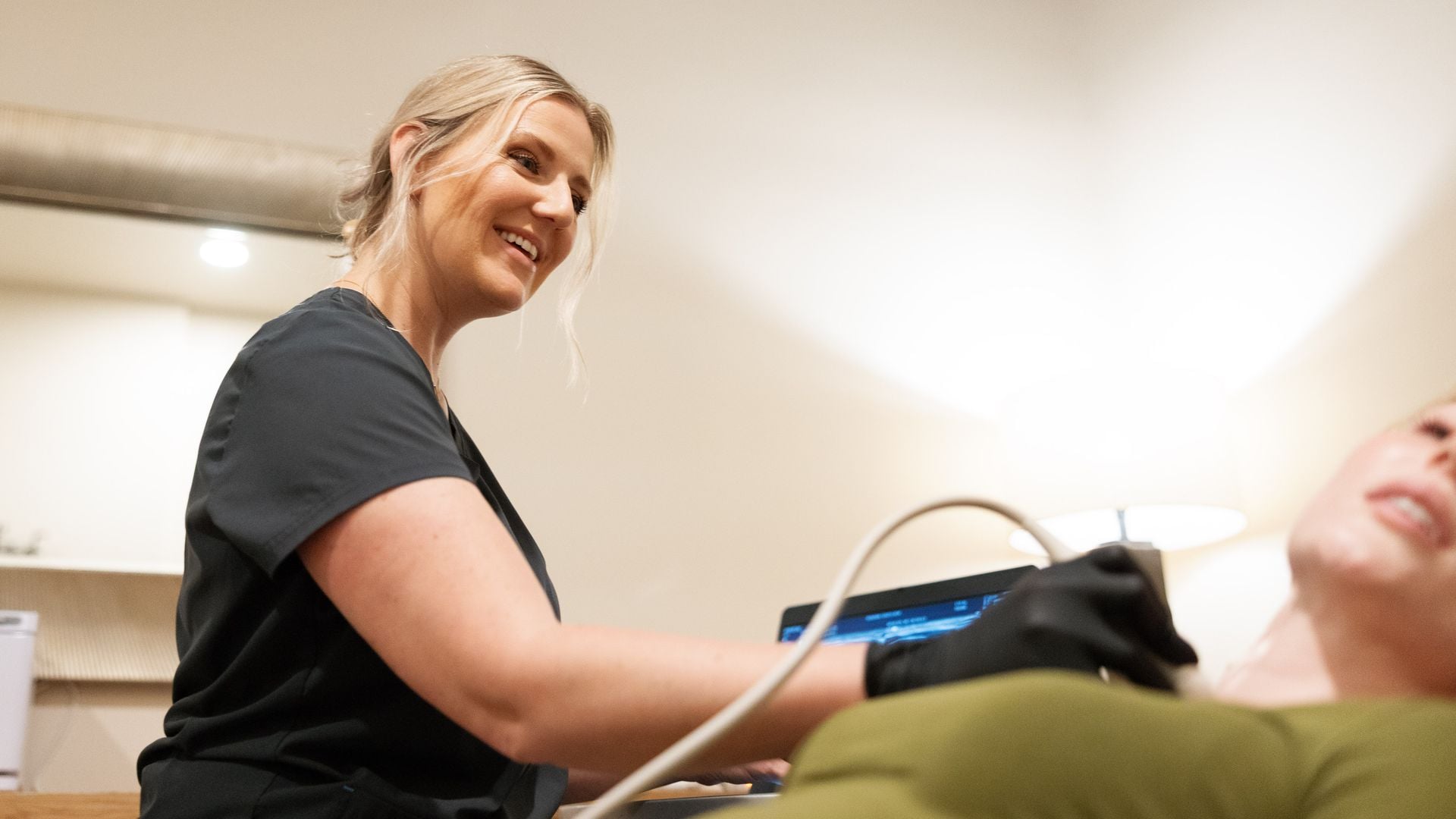
At UltraVision Imaging, pelvic ultrasounds, with or without the transvaginal portion, are performed by our registered sonographer and read by a board-certified radiologist, with the report ready within 24 hours and sent to you and, if you name one, your doctor. We see patients at our clinic, or we bring the scan to you in our California service areas. Pricing is a flat rate paid when you book, with no facility fee; HSA and FSA cards are accepted. Check availability on our booking page, and see what happens during a pelvic ultrasound and how to prepare. Please review your results with your doctor, who can put them in the context of your symptoms and history.
What else gets checked when PCOS is suspected
Because irregular periods and androgen signs have other causes, PCOS is a diagnosis made after ruling them out. Your doctor may check:
- Thyroid function (TSH), since an over- or underactive thyroid disrupts cycles.
- Prolactin, a hormone that can stop ovulation when high.
- 17-hydroxyprogesterone, to rule out a form of congenital adrenal hyperplasia.
- Pregnancy, when periods stop.
- Testosterone and other androgens, to confirm high androgen levels.
Once PCOS is diagnosed, guidelines also recommend screening for its long-term health links: blood sugar (diabetes and prediabetes are more common), cholesterol, blood pressure, sleep apnea symptoms and mood, since anxiety and depression are more common in PCOS.
PCOS is more than an ovarian condition
PCOS involves insulin resistance and metabolism as much as the ovaries, which is why lifestyle changes, even modest weight loss in those with higher weight, can restore ovulation for some people. Treatment is chosen around your goals:
| Goal | Common approaches |
|---|---|
| Regular periods and protecting the lining | Combined birth control, cyclic progestin or a hormonal IUD |
| Acne and excess hair | Birth control, spironolactone, cosmetic treatments |
| Metabolic health | Lifestyle changes, metformin, and in some cases newer weight-management medicines |
| Pregnancy | Letrozole for ovulation induction with monitoring, then other fertility treatments if needed |
Getting an accurate PCOS ultrasound
- A transvaginal scan counts small follicles much more accurately; if it is not appropriate for you, the radiologist can use ovarian volume instead.
- Scans early in the cycle, or at any time if you have no periods, avoid confusion from a dominant follicle or corpus luteum.
- Tell the sonographer if you take hormonal birth control.
- Ask that the report state the follicle count per ovary and ovarian volume, not just “polycystic appearance.”
A clear report makes it easier for your doctor to apply the current criteria and compare future scans.
PCOS across life stages
- Teens: diagnosis relies on irregular cycles and androgen signs; ultrasound is avoided because many follicles are normal at this age.
- Reproductive years: focus on cycle regulation, skin and hair symptoms, metabolic health and fertility.
- Pregnancy: PCOS raises the chance of gestational diabetes and high blood pressure, so screening is often earlier.
- Perimenopause and beyond: cycles may become more regular, but metabolic and heart health remain priorities.
Ultrasound plays its biggest role in adults, when the follicle count and ovarian volume help confirm the diagnosis and the lining is checked during irregular cycles.
Questions to ask your doctor
- Which of the three PCOS criteria do I meet?
- Was my ultrasound done with current follicle-count thresholds?
- Do I need blood sugar, cholesterol or thyroid tests?
- How should I protect my lining if my periods are infrequent?
- What are my options if I want to get pregnant?
Finally, remember that PCOS is a long-term condition that changes with life stage. A diagnosis is the beginning of a plan, and regular check-ins with your clinician help keep symptoms, metabolic health and fertility goals on track.
Frequently asked questions
Can you have PCOS without cysts on ultrasound?
Yes. PCOS is diagnosed with two of three features: irregular ovulation, signs of high androgens, and polycystic ovaries on ultrasound or a high AMH. A normal scan does not rule it out.
How many follicles count as PCOS?
Under the 2023 international guideline, 20 or more follicles of 2 to 9 mm in at least one ovary, or an ovarian volume of 10 mL or more.
What is the string of pearls sign?
Small follicles arranged around the edge of the ovary on ultrasound, a classic appearance in polycystic ovaries.
Can AMH replace an ultrasound for PCOS?
In adults, the 2023 guideline allows a high AMH instead of ultrasound. It should not be used in adolescents, and there is no single universal cutoff.
Does birth control affect a PCOS ultrasound?
Yes. It can lower the follicle count and ovarian volume. Talk to your doctor about timing before a diagnostic scan.
Should a transvaginal scan be used for PCOS?
When appropriate, yes. It counts small follicles far more accurately than a transabdominal scan.
Getting a scan with us: our pelvic and women’s health ultrasound page explains what the scan covers and how to prepare. You can book online for our clinic, or we bring the scan to you at home. A board-certified radiologist reads every study, and the report is ready within 24 hours. Your own doctor interprets the results for you.
Sources
This article is general health information, not medical advice. It does not replace a conversation with your own physician or fertility specialist, who knows your history and can interpret results in context. If you have symptoms that worry you, contact your doctor; if you think you are having a medical emergency, call 911.