
Your doctor mentioned a possible kidney stone. Or a routine scan turned up a “renal cyst” and nobody explained what that means. Or you have a dull ache in your side, blood in your urine, or a lab result showing your kidneys are not filtering as well as they should. In all of these situations, a kidney ultrasound is usually the first imaging test, and for good reason: it is quick, painless, uses no radiation and no contrast dye, and answers most of the first questions about the kidneys and bladder.
This guide explains what a kidney (renal) ultrasound can and cannot show, what words like hydronephrosis, simple cyst and echogenic mean on a report, how it compares with a CT scan for stones, how to prepare, and when symptoms mean you should skip the scheduled scan and go to an emergency room.
What a kidney ultrasound looks at
A renal ultrasound images both kidneys and, in most studies, the bladder. The sonographer measures each kidney, looks at the outer tissue (the cortex, where filtering happens) and the inner collecting system (where urine gathers before draining down the ureter), and checks the bladder before and sometimes after you empty it. Color Doppler, the part of the exam that shows blood flow in color, can be added to look at the blood vessels in and around the kidneys.
In a typical adult, each kidney measures about 10 to 12 centimeters long. The two sides are usually within a centimeter or two of each other. A kidney that is noticeably small, or two kidneys of very different size, is a clue the radiologist will mention.
The most common reasons people need one
- Flank or side pain that might be a kidney stone
- Blood in the urine, visible or found on a urine test
- Repeated urinary tract infections, especially in men or with fever
- Abnormal kidney blood tests, such as a rising creatinine or falling eGFR
- A cyst or “spot” on a previous scan that needs follow-up
- Trouble emptying the bladder, which can back urine up toward the kidneys
- High blood pressure at a young age, where the kidneys can be part of the cause
Kidney stones: what ultrasound sees, and what it can miss
Stones show up on ultrasound as bright white spots that cast a dark shadow behind them, much like a rock blocking light. Ultrasound is good at finding stones inside the kidney and at the point where the ureter enters the bladder. It is weaker in the middle stretch of the ureter, which runs deep behind the bowel, and with very small stones.
Size matters. In one emergency-department study of patients whose stones were confirmed by CT, ultrasound picked up the stone or its effects in about 90 percent of patients with stones 6 millimeters or larger, but in about 75 percent of patients with smaller stones. The good news is that many stones under 5 millimeters pass on their own, and ultrasound is very good at spotting the problem that matters most: whether a stone is blocking urine flow.
CT remains the most sensitive test for small stones, but it uses radiation, and people with stones often get scanned again and again over the years. For that reason, many guidelines suggest ultrasound first for pregnant patients, children and young adults, and for people with a known history of stones who need a check, saving CT for when ultrasound does not answer the question.
Hydronephrosis: when the kidney is swollen with urine
Hydronephrosis means the collecting system of the kidney is stretched by urine that is not draining normally. On ultrasound it looks like a dark, fluid-filled branching shape in the middle of the kidney. Radiologists usually grade it:
| Grade | What it means |
|---|---|
| Mild | Slight widening of the collecting system. Often seen with a small stone, a full bladder, or pregnancy, and can be temporary. |
| Moderate | Clear swelling of the collecting system. The cause, such as a stone or narrowing, needs to be found. |
| Severe | Marked swelling with thinning of the kidney tissue. Needs prompt attention from a doctor. |
Hydronephrosis is a finding, not a diagnosis. Common causes include a stone, an enlarged prostate, a narrowing where the kidney meets the ureter, and pressure from pregnancy. Mild hydronephrosis in pregnancy, especially on the right side, is common and usually harmless. The radiologist will describe the degree and, when visible, the likely cause; your doctor decides what to do next.
Kidney cysts: usually nothing to worry about
Simple kidney cysts are among the most common findings in all of imaging, and they become more common with age; a large share of adults over 50 have at least one. A simple cyst is a thin-walled pocket of clear fluid. On ultrasound it is black inside, round, smooth, and shows brightness behind it because sound passes easily through fluid. A cyst that meets these criteria is benign and needs no follow-up.
Radiologists use the Bosniak classification to sort cysts by how complicated they look. It was designed for CT and MRI, but ultrasound is often what first finds the cyst and tells the radiologist whether it is simple or needs a closer look.
| Bosniak category | Typical appearance | Usual next step |
|---|---|---|
| I | Simple cyst: thin smooth wall, clear fluid | No follow-up |
| II | A few thin lines inside, or tiny calcification | No follow-up |
| IIF | Slightly more complex, but not clearly concerning | Follow-up imaging over several years |
| III | Thickened walls or dividing lines that enhance with contrast | Referral to a urologist |
| IV | Solid parts inside the cyst | Referral to a urologist |
If your report says “complex cyst” or recommends CT or MRI, that is not a cancer diagnosis. It means the radiologist wants a test that can show blood flow into the cyst walls more clearly.
Other words you might see on the report
- Increased echogenicity: the kidney tissue looks brighter than normal. This can go along with chronic kidney disease and is compared with your blood tests.
- Cortical thinning: the outer filtering layer is thinner than expected, which can happen with long-standing kidney disease or high blood pressure.
- Angiomyolipoma: a very bright, well-defined spot made partly of fat. These are benign; larger ones are sometimes watched over time.
- Solid renal mass: a lump that is not fluid. This needs further imaging with CT or MRI and a urologist, because some solid masses are kidney cancers that are very treatable when found early.
- Post-void residual: how much urine stays in the bladder after you empty it. A large amount can point to a bladder outlet problem, such as an enlarged prostate.
Our guide to reading an ultrasound report explains more of the general vocabulary.
How to prepare
- Fill your bladder. Drink about 32 ounces (1 liter) of water in the hour before your scan and do not go to the bathroom. Well-hydrated kidneys and a full bladder are much easier to see. If your doctor has told you to limit fluids, follow their instructions and let us know when you book.
- You will empty your bladder partway through so the urine left behind can be measured.
- Eating is fine for a kidney and bladder study. If you are also having a full abdominal scan, follow the 8-hour fasting instructions for that one.
- Keep taking your usual medicines unless your doctor tells you otherwise.
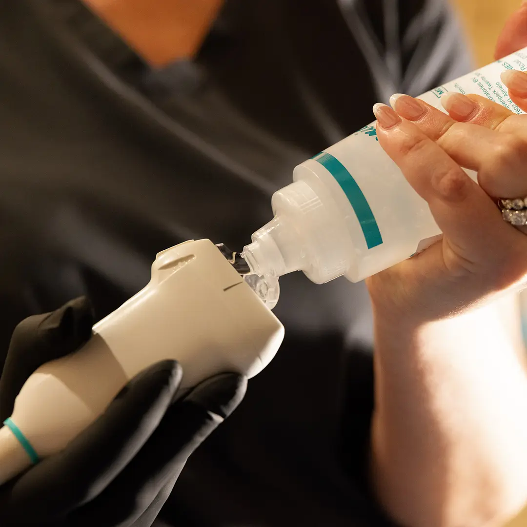
The scan itself takes about 20 to 30 minutes. You lie on your back and then on each side while the sonographer moves a gel-covered probe over your sides and lower belly. It does not hurt, though pressure over a tender kidney can be uncomfortable for a moment.
When not to wait for a scheduled scan
Go to an emergency room or call 911 if you have:
- Severe side or back pain with fever or chills, which can mean an infected, blocked kidney
- Pain so strong you cannot keep fluids down
- Inability to urinate, or very little urine output
- Heavy blood in the urine with clots or weakness
A blocked kidney with infection is a true emergency. A scheduled outpatient ultrasound is the right choice for follow-up, mild symptoms, and questions your doctor wants answered, not for this situation.
How booking works
- Enter your ZIP code on our booking page to see whether you can come to our clinic or have us bring the scan to you.
- Choose the kidney (renal) ultrasound, which includes the bladder, and pick an open time.
- Follow the prep instructions in your confirmation email. Our ultrasound prep guide explains why each step matters.
- Get your report. A board-certified radiologist reads every study and the report is ready within 24 hours. We send it to you and, if you name one, to your doctor.
Pricing is a flat rate paid when you book, with no facility fee and no surprise bill afterward. HSA and FSA cards are accepted, and we can provide a superbill if you want to submit the visit to your insurer yourself.
You do not need a referral to book a kidney ultrasound with us. Our article on getting an ultrasound without a referral explains how that works and when seeing your doctor first is the better choice. Either way, talk with your doctor about your results so they can be read alongside your symptoms and lab work.
Getting a scan with us: our abdominal ultrasound page explains what the scan covers and how to prepare. You can book online for our clinic, or we bring the scan to you at home. A board-certified radiologist reads every study, and the report is ready within 24 hours. Your own doctor interprets the results for you.
Frequently asked questions
Can an ultrasound show kidney stones?
Yes. Ultrasound shows most stones inside the kidney and near the bladder, and it is very good at showing whether a stone is blocking urine flow. It can miss small stones in the middle part of the ureter, where CT is more sensitive.
Is a kidney ultrasound as good as a CT scan?
For stones, CT finds more small stones. Ultrasound uses no radiation, needs no dye and answers most first questions, so many guidelines recommend it first for pregnant patients, young people and people with repeat stones.
What does hydronephrosis mean?
It means the inside of the kidney is stretched by urine that is not draining normally. It is usually caused by something downstream, such as a stone or an enlarged prostate. Mild hydronephrosis can be temporary.
Should I worry about a kidney cyst?
Most kidney cysts are simple fluid-filled cysts that are benign and need no follow-up. If a cyst looks complex, the radiologist may suggest CT or MRI to look at it more closely.
Do I need to fast for a kidney ultrasound?
Usually not. Drink about 32 ounces of water an hour before and arrive with a full bladder. If a full abdominal scan is being done at the same time, follow the fasting instructions for that study.
How long does a kidney ultrasound take?
About 20 to 30 minutes. A board-certified radiologist reads the images and the report is ready within 24 hours.
This article is general health information, not medical advice. It does not replace a conversation with your own physician, who knows your history and can interpret results in context. If you have symptoms that worry you, contact your doctor; if you think you are having a medical emergency, call 911.