
Thyroid nodules are among the most common findings in adult medicine. Many are found incidentally on imaging done for other reasons; some are felt on a careful neck exam during an annual physical. Most are benign. The task for the physician is to identify the minority that need a biopsy or close follow-up without sending every patient down a cascade of tests.
Ultrasound is the central tool for that decision, and the way the report is written matters as much as the scan itself. This guide covers the usual first steps after a palpable nodule, how the ACR Thyroid Imaging Reporting and Data System (TI-RADS) turns ultrasound features into a recommendation, and how a mobile thyroid study fits into concierge care.
First steps after a palpable nodule
American Thyroid Association guidance recommends measuring serum TSH and performing a diagnostic thyroid ultrasound with a survey of the cervical lymph nodes for patients with a known or suspected nodule. If TSH is low, a radionuclide scan may be considered to identify a hyperfunctioning nodule, which is rarely malignant. The history matters too: childhood head and neck radiation, a family history of thyroid cancer or certain genetic syndromes, rapid growth, hoarseness or difficulty swallowing all raise concern.
Screening ultrasound of the thyroid in people without symptoms or findings is a different matter; the USPSTF recommends against it. A palpable nodule, however, is a diagnostic question.
What the ultrasound documents
A complete thyroid study measures both lobes and the isthmus, then characterizes each nodule and surveys the lateral neck for abnormal lymph nodes. For each nodule, the report describes size in three dimensions and the features TI-RADS scores.
How ACR TI-RADS works
TI-RADS assigns points in five feature categories:
- Composition: cystic or spongiform (0), mixed cystic and solid (1), solid or almost completely solid (2).
- Echogenicity: anechoic (0), hyper- or isoechoic (1), hypoechoic (2), very hypoechoic (3).
- Shape: wider-than-tall (0), taller-than-wide (3).
- Margin: smooth or ill-defined (0), lobulated or irregular (2), extra-thyroidal extension (3).
- Echogenic foci: none or large comet-tail artifacts (0), macrocalcifications (1), peripheral rim calcifications (2), punctate echogenic foci (3).
The total places the nodule in a category from TR1 (benign) to TR5 (highly suspicious). Each category has size thresholds for fine-needle aspiration and for follow-up imaging:
- TR1 and TR2: no FNA or routine follow-up.
- TR3 (mildly suspicious): FNA if 2.5 cm or larger; follow-up if 1.5 cm or larger.
- TR4 (moderately suspicious): FNA if 1.5 cm or larger; follow-up if 1.0 cm or larger.
- TR5 (highly suspicious): FNA if 1.0 cm or larger; follow-up if 0.5 cm or larger.
Recommended follow-up intervals lengthen for lower-risk categories. The radiologist’s report applies these rules, but the ordering physician integrates them with the patient’s history, preferences and other findings. A patient with a strong family history may warrant a different approach than the table alone suggests.
Why consistency matters for follow-up
Thyroid nodules are often followed for years. Growth is judged by comparing measurements over time, so measurement technique matters. A study performed to a standard protocol, with the same caliper conventions each time, makes those comparisons reliable. That is one more reason to order a formal study rather than rely on a quick bedside look when a nodule will be followed.
Lymph nodes
A thyroid ultrasound should include the central and lateral neck. Lymph nodes with suspicious features, such as loss of the fatty hilum, rounded shape, cystic change, calcifications or abnormal blood flow, change the work-up regardless of the nodule’s TI-RADS category.
Common report findings and what they usually mean
Several phrases appear frequently in thyroid reports and can prompt questions from patients:
- Spongiform or simple cystic nodule. These are TI-RADS TR1 or TR2 patterns and are considered benign or not suspicious. They usually need no biopsy or routine follow-up.
- Colloid or comet-tail artifact. Large comet-tail artifacts within cystic areas are a reassuring feature and score zero points.
- Multinodular goiter. Several nodules in an enlarged gland. Each nodule is scored separately, and the report typically highlights up to four of the most suspicious.
- Heterogeneous echotexture. A patchy appearance of the whole gland that can accompany autoimmune thyroid disease, such as Hashimoto thyroiditis; it is interpreted alongside thyroid antibodies and function tests.
- Punctate echogenic foci. Tiny bright spots within a solid nodule that raise the score and often lower the size threshold for biopsy.
The ordering physician puts these descriptions into context with the patient’s history and labs.
Follow-up intervals
For nodules that meet follow-up but not biopsy thresholds, ACR TI-RADS suggests imaging intervals that lengthen as risk falls, with follow-up typically stopping after about five years if the nodule is stable. Significant growth is generally defined as an increase of 20% or more in at least two dimensions with a minimum increase of 2 mm, or a 50% or greater increase in volume. Consistent technique between studies matters for that comparison, which is one reason practices prefer the same provider and protocol for follow-up.
After a biopsy recommendation
If the report recommends fine-needle aspiration, the ordering physician typically refers to an endocrinologist, radiologist or surgeon who performs ultrasound-guided biopsies. Cytology is reported using the Bethesda System, which assigns each sample to one of six categories that guide next steps. Mobile diagnostic ultrasound provides the imaging that leads to that decision; the biopsy itself is performed in a procedural setting.
Talking with patients about nodules
Patients often hear “nodule” and think “cancer.” It helps to tell them, before the study, that nodules are very common, that most are benign, and that the purpose of the ultrasound is to decide whether anything more needs to be done. Patients who understand the plan in advance are calmer when the report arrives.
When ultrasound is not the whole answer
Ultrasound is the primary imaging test for thyroid nodules, but a few situations call for something else. A large goiter extending behind the breastbone may need CT to assess its full extent. A low TSH points toward a functioning nodule, which a radionuclide scan can confirm. Suspicious lymph nodes may prompt biopsy of the node itself. The radiologist’s report often notes when another study would add information, and the ordering physician decides on next steps.
Thyroid imaging in pregnancy and in younger patients
Ultrasound uses no radiation, so it is safe during pregnancy and preferred for younger patients who may need years of follow-up. Nodules found during pregnancy are generally evaluated with ultrasound in the same way, and the timing of any biopsy is a decision for the treating physician and the patient. For adolescents, thyroid nodules carry a somewhat higher chance of malignancy than in adults, and pediatric-specific guidance applies; these patients are usually managed with a pediatric endocrinologist.
A mobile thyroid study
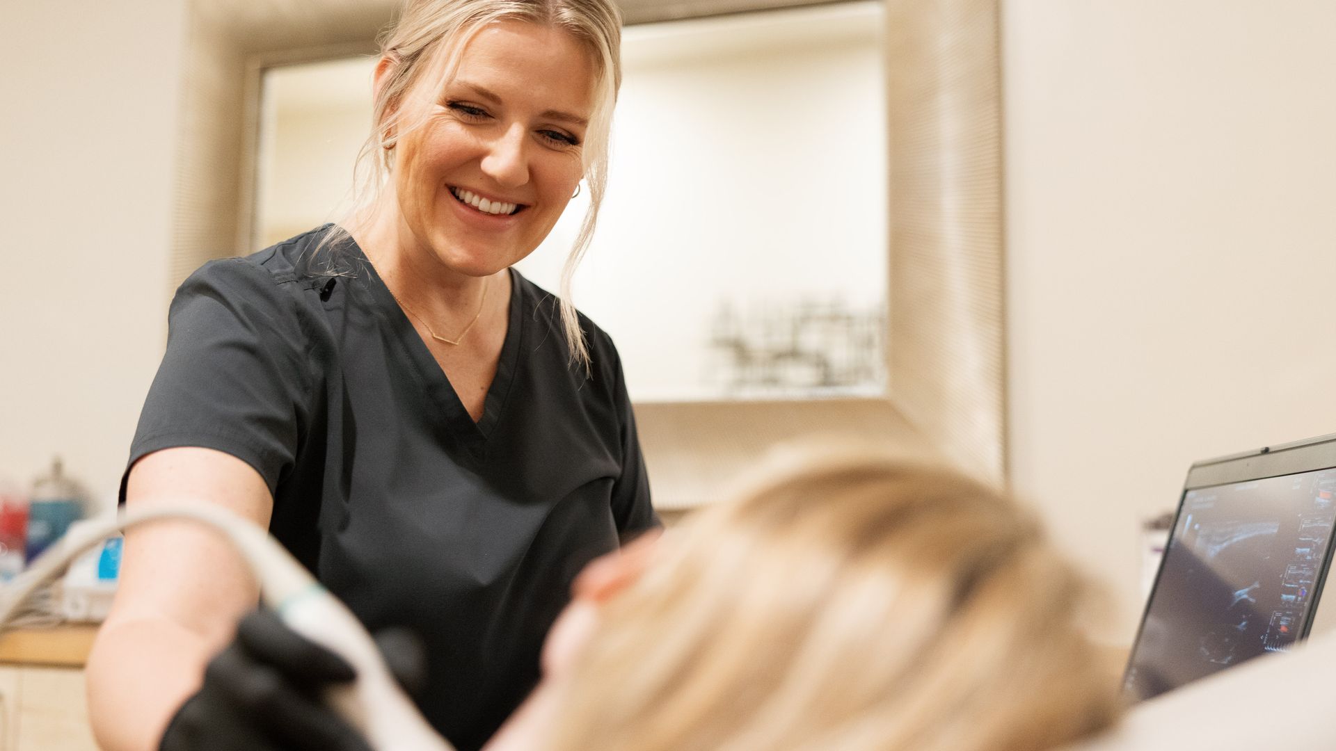
Thyroid ultrasound is well suited to a home or office visit. It needs no preparation, takes about 20 to 30 minutes, and requires only a surface to lie on with the neck slightly extended. For executive and concierge patients, a follow-up thyroid study can be scheduled at their office between meetings.
UltraVision Imaging performs diagnostic thyroid and neck ultrasound with lymph node survey for concierge, house-call and private-medicine practices across Los Angeles, San Diego County, Orange County and Palm Springs, the Bay Area and the Sacramento region. A board-certified radiologist reads every study, and the report goes to the ordering physician. See every region on our page for physicians. Our team will be in touch to coordinate scheduling.
Frequently asked questions
What should be ordered first for a palpable thyroid nodule?
Guidance from the American Thyroid Association recommends a serum TSH and a diagnostic thyroid ultrasound with a lymph node survey.
What is ACR TI-RADS?
A system that scores five ultrasound features of a thyroid nodule and assigns a category from TR1 to TR5 with size thresholds for biopsy and follow-up.
Which TI-RADS categories need a biopsy?
It depends on size: TR3 at 2.5 cm or more, TR4 at 1.5 cm or more, TR5 at 1.0 cm or more. The ordering physician makes the final decision.
Should healthy adults be screened with thyroid ultrasound?
The USPSTF recommends against thyroid cancer screening in asymptomatic adults. A palpable nodule is a diagnostic question, not screening.
Does a thyroid ultrasound need preparation?
No. The patient lies with the neck slightly extended; the study takes about 20 to 30 minutes.
Are lymph nodes checked?
Yes. A complete study surveys the central and lateral neck for abnormal lymph nodes.
Sources
- Tessler FN et al. ACR Thyroid Imaging, Reporting and Data System (TI-RADS): White Paper of the ACR TI-RADS Committee. J Am Coll Radiol 2017
- Haugen BR et al. 2015 American Thyroid Association management guidelines for adult patients with thyroid nodules and differentiated thyroid cancer. Thyroid 2016
- USPSTF: Thyroid cancer screening (2017)
- RadiologyInfo.org (ACR and RSNA): Thyroid ultrasound
This article is general information for licensed clinicians and practice staff. It is not medical or legal advice and does not replace clinical judgment. UltraVision Imaging performs diagnostic ultrasound; a board-certified radiologist interprets every diagnostic study, and the ordering physician directs care.