
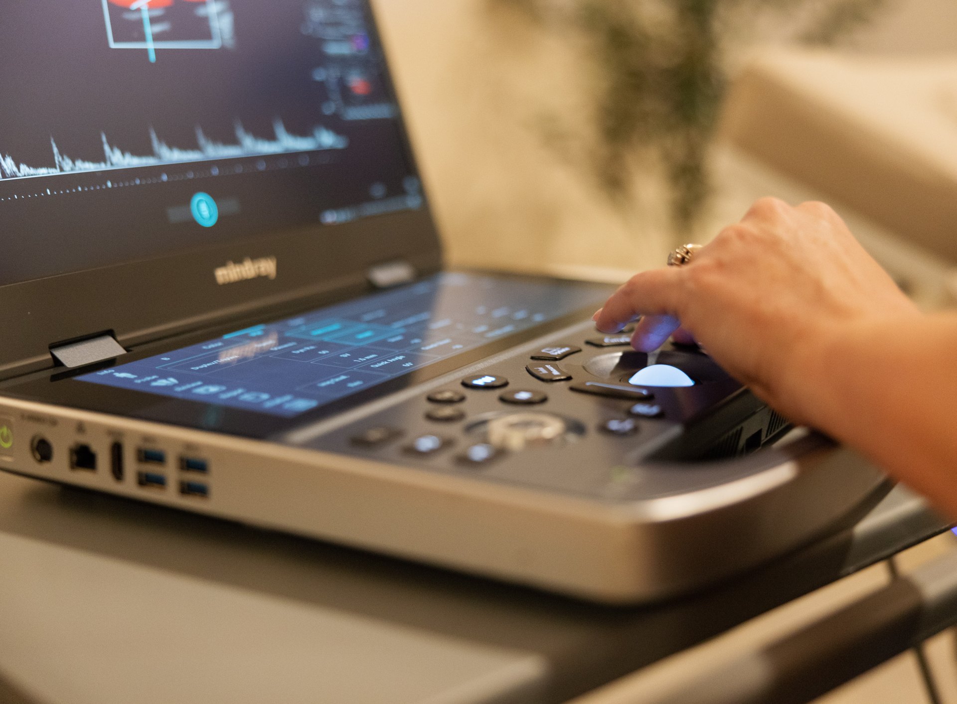
“Is ultrasound safe?” is one of the most common questions people ask before a scan, and it is a good one. Most people have heard that X-rays and CT scans involve radiation, and it is natural to wonder whether ultrasound does too. The short answer from the U.S. Food and Drug Administration is that ultrasound imaging uses sound waves, not ionizing radiation, and has a long record of safe use. The longer answer is more interesting: ultrasound is energy, energy can interact with tissue, and the professional organizations that oversee ultrasound ask that it be used thoughtfully, for a medical reason, by trained people. This article explains what that means in plain language.
One thing to say up front: this is general information about how ultrasound works and how its safety is managed. Whether a particular scan is right for you, and how often you should have one, are questions for your own doctor, who knows your history and the reason for the test.
Does ultrasound use radiation?
Not the kind people usually worry about. X-rays, CT scans, mammograms and nuclear medicine studies use ionizing radiation, which carries enough energy to knock electrons off atoms and can damage DNA. That is why radiation dose is tracked carefully for those tests.
Ultrasound works differently. A handheld transducer sends high-frequency sound waves, far above the range of human hearing, into the body. The waves bounce off tissues and fluid, the transducer picks up the echoes, and a computer turns them into a moving image. The FDA describes ultrasound as involving no ionizing radiation exposure. You may occasionally see ultrasound grouped under “radiation-emitting products” on FDA web pages; that is a regulatory category that includes many kinds of energy, and it does not mean ultrasound exposes you to X-ray-type radiation.
| Imaging test | Uses ionizing radiation? | How it makes images |
|---|---|---|
| Ultrasound | No | Sound-wave echoes |
| MRI | No | Strong magnetic field and radio waves |
| X-ray | Yes | X-ray beam through the body |
| CT scan | Yes | Many X-ray images combined by computer |
| Mammogram | Yes (low dose) | Specialized low-dose X-ray of the breast |
| Nuclear medicine and PET | Yes | Small amount of radioactive tracer |
Because there is no ionizing radiation, ultrasound is often the first imaging test for children, for people who are pregnant and for many abdominal and pelvic questions. Whether it is the right test for a particular problem is a judgment your doctor makes based on what they need to see; ultrasound cannot see through bone or gas well, so sometimes a different test is the better choice.
If not radiation, what are the possible effects?
The FDA notes two ways ultrasound energy can affect tissue, and the professional literature uses two numbers on the screen to keep track of them.
Heating and the thermal index (TI)
As sound waves pass through tissue, a small amount of the energy is absorbed and turned into heat. In everyday diagnostic imaging, any warming is slight. The thermal index, shown on modern ultrasound screens, is an estimate of how much the tissue could warm under the current settings. Higher-output modes, such as spectral Doppler used to measure blood flow, can raise it. The American Institute of Ultrasound in Medicine (AIUM) asks operators to watch the TI and keep it low, particularly in early pregnancy; for obstetric scans it advises keeping the TI at or below 0.7 when adequate images are possible and not prolonging the exam.
Mechanical effects and the mechanical index (MI)
Sound waves create tiny pressure changes. In tissue that contains gas, these changes can, in principle, cause small gas bubbles to form or vibrate, an effect called cavitation. The mechanical index estimates the likelihood of these effects. The FDA notes that the long-term effects of tissue heating and cavitation are not known, which is part of why caution is built into how ultrasound is used, even though no harm from diagnostic imaging has been proven in humans.
How output is limited
FDA guidance for manufacturers of diagnostic ultrasound systems sets upper limits on acoustic output. For systems that display TI and MI on screen, the guidance caps the derated spatial-peak temporal-average intensity at 720 mW/cm² and the mechanical index at 1.9 (with lower limits for eye exams). These limits are for the equipment; within them, the person scanning chooses the settings, which is where training matters.
| Term | What it means | Who watches it |
|---|---|---|
| Thermal index (TI) | Estimate of possible tissue warming | The sonographer, on the screen during the scan |
| Mechanical index (MI) | Estimate of possible mechanical effects such as cavitation | The sonographer, on the screen during the scan |
| ALARA | “As low as reasonably achievable”: the lowest output and shortest time that still gives a good image | Everyone performing ultrasound |
| Acoustic output limits | Maximum output allowed for the machine | The manufacturer, under FDA guidance |
If you are curious about these numbers, you are welcome to ask your sonographer to point them out. What they mean for your own health, if anything, is a question for your doctor.
ALARA: why sonographers use the lowest effective settings
ALARA stands for “as low as reasonably achievable.” Both the FDA and the AIUM ask everyone who performs ultrasound to follow it. In practice it means three things: start at a low output and raise it only if the image needs it, use higher-output modes such as Doppler only when they answer a clinical question, and keep the scan as short as possible while still getting a complete, diagnostic study.
The AIUM’s October 2026 statement on the appropriate use of medical ultrasound repeats these points. It says ultrasound should be performed by appropriately licensed or credentialed professionals working within their scope of practice, that studies should be supervised and interpreted by a qualified licensed provider, and that operators should use the lowest output and shortest exam time needed. It also says the standards should not change based on where the scan happens. That last point matters for scans done in a home: the same training, the same protocol, the same equipment standards and the same physician interpretation apply.
Is ultrasound safe during pregnancy?
Ultrasound is the most widely used imaging method for looking at a developing baby, and it is the imaging test obstetric care relies on. The American College of Obstetricians and Gynecologists (ACOG) states that ultrasonography and MRI are not associated with risk and are the imaging techniques of choice in pregnancy, while also saying they should be used prudently and only when expected to answer a relevant clinical question or otherwise benefit the patient.
The AIUM’s 2020 statement on prudent use in pregnancy takes a similarly careful line. It concludes that there is insufficient evidence that diagnostic ultrasound causes recognized harm in humans, notes that much of the research predates newer machine output levels, and therefore recommends a conservative approach: get the information that is needed at the lowest exposure. It also advises extra caution with spectral Doppler in the first trimester, using it only when the benefit clearly outweighs the risk and keeping the TI and exam time low.
Your OB, midwife or other prenatal care provider decides which scans you need and when. Our pregnancy ultrasound timeline explains the scans commonly offered at each stage, and your prenatal care team can tell you which apply to you.
What the FDA and AIUM say about keepsake and non-medical scans
Both organizations have addressed ultrasound done without a medical reason, and it is fair to share their positions plainly.
- The FDA discourages using ultrasound for non-medical purposes, such as fetal “keepsake” videos. It considers keepsake images acceptable when they come from a medically indicated exam with no additional exposure.
- The AIUM’s 2020 statement strongly discourages non-medical fetal scanning, meaning scans only to view the fetus, obtain images or learn fetal sex without a medical reason, and its 2026 statement says it strongly discourages the non-medical use of ultrasound during pregnancy.
- Both groups also caution about over-the-counter fetal heart monitors used without training.
We offer an elective heartbeat and gender visit. It is non-diagnostic: it does not include a radiologist’s report and it does not replace any of the scans your OB or midwife orders. If you are considering an elective visit, it is reasonable to read these positions and talk with your prenatal care provider first. The decision is yours to make with them.
How often is it safe to have an ultrasound?
There is no fixed lifetime limit, because there is no cumulative radiation dose to track. That does not mean “more is always better.” The professional view is that every scan should have a reason. Some people have frequent scans for good medical reasons: follicle tracking during fertility treatment may involve several transvaginal scans within a cycle, and some pregnancies call for growth scans every few weeks. Others may have a single scan in years. How often you should be scanned depends on why, and that is a decision for your doctor or care team.
Two practical points also apply to scan frequency:
- Repeat scans can find incidental findings, small things unrelated to the original question, that may lead to more tests. That is not a safety issue in the radiation sense, but it is worth discussing with your doctor before choosing a scan that is not medically needed.
- Follow-up intervals in a report, such as “repeat in six months,” are recommendations for your physician to consider in context. Our post on what your ultrasound report means explains how reports are laid out; your doctor explains what any recommendation means for you.
Other everyday safety questions
Is the gel safe?
Ultrasound gel is a water-based gel that helps sound pass from the transducer into the skin. It wipes off easily. If your skin is sensitive to any products, mention it before the scan.
Is a transvaginal scan safe?
The transvaginal probe uses the same kind of sound energy, placed closer to the pelvic organs for a clearer picture. It is covered with a protective cover for each patient. The transvaginal portion is always your choice, and you can ask to stop at any time. Our post on what happens during a pelvic ultrasound walks through the exam.
What about implants, pacemakers or metal?
Unlike MRI, ultrasound does not use a magnetic field, so pacemakers, joint replacements and other metal implants are not a safety concern for the scan. Tell the sonographer about them anyway, since they can affect what the images show.
Why diagnostic scans are medically supervised
Safety is not only about energy. A scan that is performed poorly, read by someone without the right training or never reviewed by a physician can cause harm in a different way, through a missed finding or false reassurance. That is why diagnostic ultrasound has three distinct roles. Our sonographer performs the scan and captures the images. A board-certified radiologist reads every study and writes the report, which is ready within 24 hours. Your own doctor interprets that report for you, in the context of your history, exam and labs. We never diagnose, and the sonographer will not tell you what they see; our post Why won’t the sonographer tell me what they see? explains why.
When to seek urgent care instead of a scheduled scan
An outpatient ultrasound is not a substitute for urgent care. If you are pregnant and have heavy bleeding, severe abdominal pain, fainting, a severe headache with vision changes, or a sudden gush of fluid, contact your OB right away or call 911. For anyone, call 911 for chest pain, trouble breathing, signs of stroke, or severe pain that came on suddenly. We do not provide emergency or after-hours reads.
Getting your scan with us
You can book most scans online without a referral; DVT (vein clot) studies and the second-trimester anatomy scan require a referral, which you upload in the booking form so we can confirm it before your visit. The form asks for the name of the doctor who should receive the report (type “None” if you do not have one). Payment is a flat rate paid when you book, with no facility fee, and HSA/FSA cards are accepted. We see patients at our clinic and also bring the scan to you at home across our California service areas. Read how to prepare for an ultrasound for prep by scan type, then book your appointment.
Sources
- U.S. FDA: Ultrasound Imaging
- U.S. FDA: Marketing Clearance of Diagnostic Ultrasound Systems and Transducers, guidance (2023)
- AIUM: Prudent Use and Safety of Diagnostic Ultrasound in Pregnancy (2020)
- AIUM: Statement on the Appropriate Use of Medical Ultrasound (October 2026)
- ACOG Committee Opinion 723: Guidelines for Diagnostic Imaging During Pregnancy and Lactation
- Safety of Obstetric Ultrasound: Mechanical and Thermal Indexes, a Systematic Review (PMC)
- Health Canada: Guidelines for the Safe Use of Diagnostic Ultrasound
- RadiologyInfo (ACR/RSNA): General Ultrasound
Frequently asked questions
Does ultrasound use radiation?
Ultrasound uses high-frequency sound waves, not ionizing radiation. The FDA describes it as involving no ionizing radiation exposure, unlike X-rays, CT scans and mammograms.
Can ultrasound heat tissue?
Ultrasound energy can warm tissue slightly, and it can cause small mechanical effects in tissue containing gas. Modern machines display a thermal index and a mechanical index, and sonographers follow the ALARA principle to keep output and scan time as low as reasonably achievable.
Is ultrasound safe in pregnancy?
ACOG states that ultrasonography and MRI are not associated with risk and are the imaging techniques of choice in pregnancy, while advising that they be used prudently for a clinical reason. The AIUM recommends a conservative approach with the lowest exposure needed. Your prenatal care provider decides which scans you need.
How many ultrasounds can I safely have?
There is no fixed lifetime limit because there is no cumulative radiation dose. Professional groups say each scan should have a medical reason. How often you should be scanned depends on why, and that is a decision for your doctor.
What do the FDA and AIUM say about keepsake ultrasounds?
The FDA discourages non-medical uses such as fetal keepsake videos, and the AIUM strongly discourages non-medical fetal ultrasound. Our elective heartbeat and gender visit is non-diagnostic, has no radiologist report and does not replace scans your OB orders. Talk with your prenatal care provider before choosing one.
Is ultrasound safe with a pacemaker or metal implant?
Ultrasound does not use a magnetic field, so pacemakers and metal implants are not a safety concern for the scan itself. Let the sonographer know about them because they can affect the images.
This article is general health information, not medical advice. It does not replace a conversation with your own physician, who knows your history and can interpret results in context. If you have symptoms that worry you, contact your doctor; if you think you are having a medical emergency, call 911.