
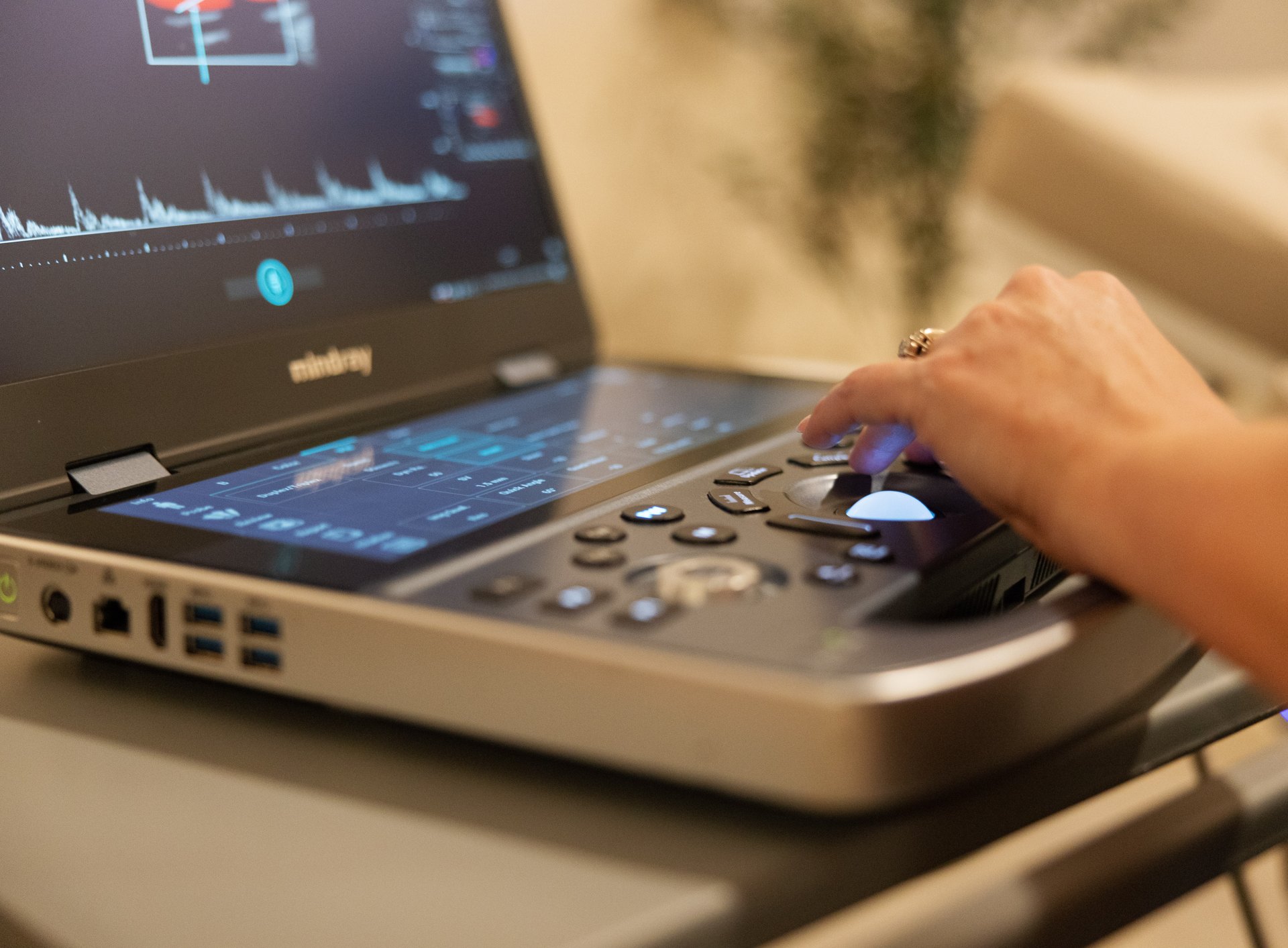
A swollen, aching calf after a long flight. A leg that has felt tight and warm since surgery. An arm that puffed up around an IV line. These are the situations where doctors worry about a deep vein thrombosis (DVT), a blood clot in one of the deep veins, and where a venous duplex ultrasound is the standard test to find out.
This guide explains when a clot is likely, what the ultrasound involves, what the different kinds of DVT exam can and cannot show, what the results mean, and the symptoms that mean you should call 911 instead of booking anything.
Call 911 if you have these symptoms
A clot that breaks loose and travels to the lungs is a pulmonary embolism, which can be life-threatening. Call 911 or go to an emergency room right away if you have:
- Sudden shortness of breath
- Chest pain that is sharp or worse when you breathe in
- Coughing up blood
- A racing heartbeat, fainting or feeling faint
These symptoms need emergency care, not a scheduled ultrasound.
Signs a leg or arm clot may be present
- Swelling of one leg or arm, especially if it is new and one-sided
- Pain, cramping or tenderness, often in the calf or thigh
- Warmth over the area
- Redness or a darker skin color
- Visible surface veins that were not there before
Some clots cause few or no symptoms. Others that look like clots turn out to be a pulled muscle, a burst cyst behind the knee (a Baker cyst), or skin infection. That is why imaging matters: treatment with blood thinners has real risks, so doctors want to be sure.
Who is at higher risk
- Recent surgery, especially hip, knee or abdominal surgery
- Long periods without moving: hospital stays, bed rest, long flights or car rides
- Pregnancy and the first weeks after giving birth
- Estrogen-containing birth control or hormone therapy
- Cancer and some cancer treatments
- A previous clot, or a family history of clots
- A central line, PICC line or pacemaker wires, which raise the risk of arm clots
- Smoking, obesity and older age
How doctors decide who needs the scan
Doctors often start with a scoring checklist called the Wells score, which adds up risk factors and symptoms, and sometimes a blood test called a D-dimer. A normal D-dimer in someone at low risk can rule out a clot without imaging. A higher score or a positive D-dimer usually leads to an ultrasound. This is one reason we ask for a referral for DVT studies: your doctor’s assessment shapes how the results are used.
What the ultrasound actually does
The main technique is compression. A healthy vein flattens completely when the sonographer presses on it with the probe. A vein with a clot inside will not collapse. The sonographer works along the deep veins, pressing every couple of centimeters, and uses color Doppler to watch blood flow and listen to how it changes when you breathe or when your calf is squeezed.
There are two common approaches for legs:
| Exam | What it covers | Strength | Limitation |
|---|---|---|---|
| Two-point (limited) compression | The groin vein and the vein behind the knee | Quick and highly reliable for the larger clots that matter most | Does not check calf veins; a repeat scan in about a week is often advised if the first is normal |
| Whole-leg (complete) duplex | The deep veins from the groin to the ankle, including the calf | Answers the question in one visit | Takes longer and finds small calf clots that may or may not need treatment |
In a large randomized trial of more than 2,000 outpatients published in JAMA, both strategies proved equally safe: over the following three months, about 1% of people sent home after a normal result went on to have a clot, whichever exam they had. Your doctor and the radiologist decide which approach fits your situation.
Arm studies follow the same idea, checking the veins of the arm, the armpit and the base of the neck. Part of the vein under the collarbone cannot be compressed, so the sonographer relies more on flow patterns there.
What the exam feels like
- You lie on your back, with your leg slightly turned out and your head raised a little, which helps the leg veins fill.
- The sonographer presses firmly with the probe. Over a tender, swollen area this can be uncomfortable, but it is brief.
- A one-leg study usually takes 20 to 40 minutes; both legs take longer.
- No preparation is needed. Wear loose clothing that is easy to move out of the way.
Understanding the results
- No DVT: the deep veins compressed fully with normal flow. If symptoms continue or worsen, your doctor may repeat the scan in about a week, especially after a two-point exam.
- Acute DVT: a fresh clot. The vein is usually widened and does not compress. This needs your doctor today, because treatment usually starts right away.
- Chronic or old clot: scarring from a previous clot, with a thickened vein wall. Comparing with any earlier scan is very helpful.
- Superficial vein clot (superficial thrombophlebitis): a clot in a vein near the skin. Usually less serious, but one near the groin can extend into the deep system, so your doctor will decide on treatment.
- Other findings: a Baker cyst, fluid in the muscle, or swollen lymph nodes can explain symptoms when no clot is present.
How booking a DVT study works with us
Because a clot can need treatment the same day, we handle DVT studies a little differently from our other scans:
- A referral is required. It means the result goes straight to the doctor who will act on it. See our article on getting an ultrasound with or without a referral.
- Call us to book at (916) 751-1944 so we can find the soonest time and confirm the right exam. You can also book online from our booking page.
- A board-certified radiologist reads every study, and the report is ready within 24 hours and sent to your doctor.
If you think you have a clot and cannot be seen soon, do not wait for an appointment: go to urgent care or an emergency room. Pricing for our scheduled studies is a flat rate paid when you book, and HSA and FSA cards are accepted. Please review your results with your doctor, who will decide on any treatment.
Reducing your risk of a clot
- On long flights or car rides, get up and walk every hour or two, and flex your calves while seated.
- Stay hydrated and avoid sitting with your legs crossed for long periods.
- After surgery, follow your team’s plan for early walking, compression stockings or blood-thinning medicine.
- If you take estrogen-containing birth control or hormone therapy and have other risk factors, review them with your doctor.
- Know the warning signs if you have had a clot before.
After a DVT is found
Most people with a leg DVT are treated with blood-thinning tablets at home for at least three months. Your doctor may repeat the ultrasound in some situations, for example if symptoms worsen, or to establish a new baseline before stopping treatment, which helps if symptoms return later. Some people develop long-term swelling or aching after a clot; compression and exercise can help.
If you have had a clot before, keep a copy of your last ultrasound report; comparing old and new scans helps tell a new clot from old scarring.
Getting a scan with us: our vascular ultrasound page explains what the scan covers and how to prepare. You can book online for our clinic, or we bring the scan to you at home. A board-certified radiologist reads every study, and the report is ready within 24 hours. Your own doctor interprets the results for you.
Frequently asked questions
How is a DVT diagnosed?
Usually with a venous duplex ultrasound. The sonographer presses on the deep veins with the probe; a healthy vein collapses, while a vein with a clot does not. Doctors often use a risk score and a D-dimer blood test to decide who needs the scan.
Does a DVT ultrasound hurt?
It is not painful, but firm pressure over a tender, swollen area can be uncomfortable for a moment. A one-leg exam usually takes 20 to 40 minutes.
What is the difference between a two-point and a whole-leg DVT scan?
A two-point exam checks the groin vein and the vein behind the knee and may need a repeat scan in about a week. A whole-leg exam checks the deep veins down to the ankle in one visit. Both proved equally safe in a large clinical trial.
When should I go to the ER instead?
Call 911 for sudden shortness of breath, chest pain, coughing up blood, or fainting, which can mean a clot has reached the lungs.
Do I need a referral for a DVT ultrasound?
For DVT studies we require a referral so the result goes straight to the doctor who will treat you. Call (916) 751-1944 to book.
Do I need to prepare for a DVT ultrasound?
No. Wear loose clothing and take your usual medicines unless your doctor tells you otherwise.
This article is general health information, not medical advice. It does not replace a conversation with your own physician, who knows your history and can interpret results in context. If you have symptoms that worry you, contact your doctor; if you think you are having a medical emergency, call 911.