
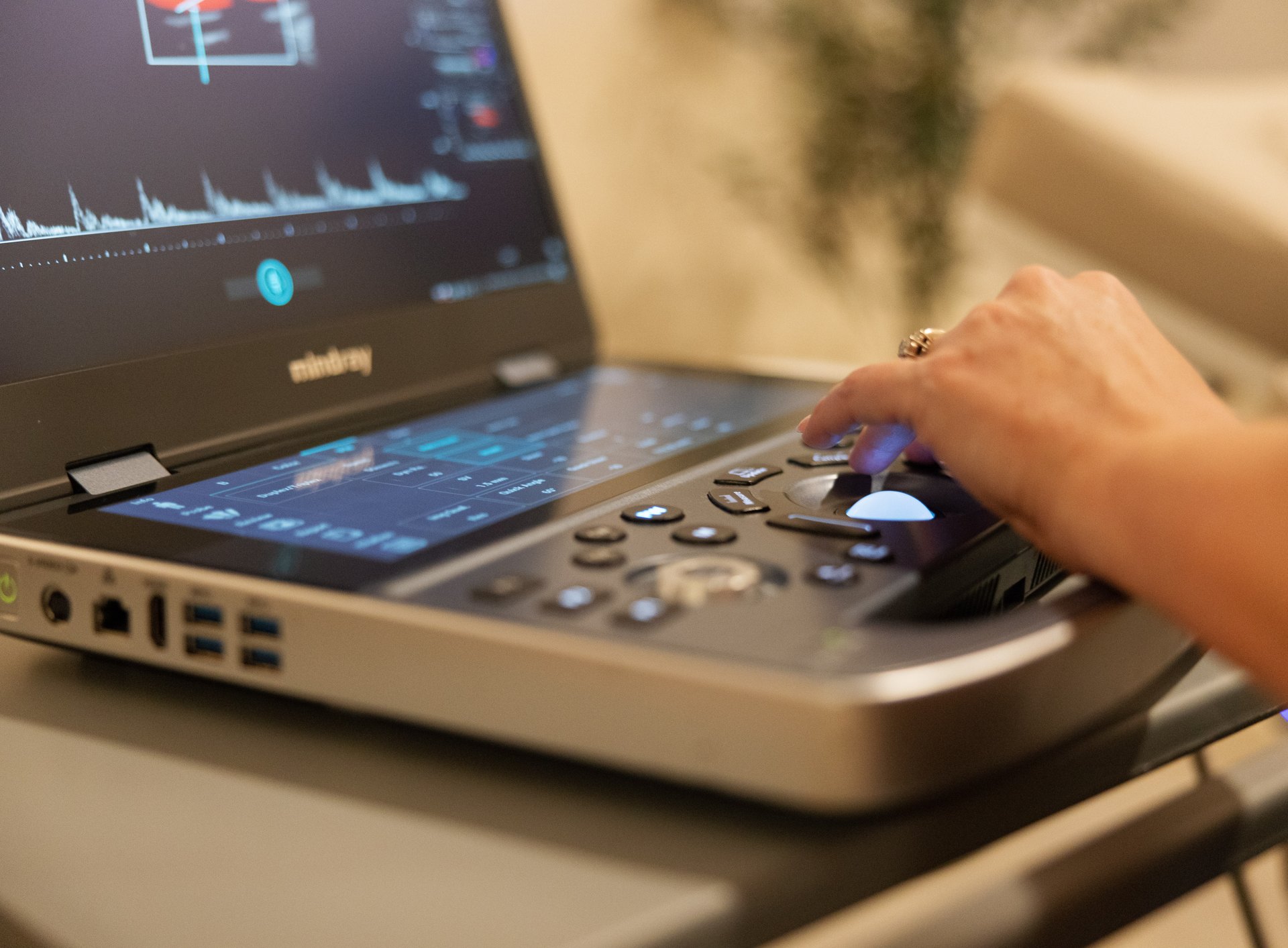
Someone finds a lump. Someone else has a vague pain that won’t go away, or a family history that keeps them up at night. The question that follows is almost always the same: Can an ultrasound tell me whether this is cancer? And right behind it: Or do I need a CT or an MRI?
The honest answer has three parts. Ultrasound is very good at some things, genuinely limited at others, and almost never the test that gives a final yes-or-no answer about cancer by itself. This article explains what ultrasound can show, what it cannot, where it is the right first test, and when CT or MRI does a better job.
The short answer
Ultrasound can find many abnormalities, characterize them (solid or fluid, size, shape, borders, blood flow) and, for several organs, sort them into categories that tell your doctor whether anything further is needed. What ultrasound cannot do is prove that something is or is not cancer. That takes tissue, which means a biopsy. As the American Cancer Society puts it, “an ultrasound might not be able to tell whether a tumor is cancer.”
That is not a weakness unique to ultrasound; CT and MRI have the same limit. All imaging describes; pathology diagnoses. The value of imaging is in deciding, quickly and without a needle, whether a biopsy is even worth discussing.
What ultrasound does well
Solid versus fluid
This is ultrasound’s superpower. Fluid returns no echoes and appears black; solid tissue returns echoes and appears gray. A “lump” that turns out to be a simple cyst (a thin-walled sac of fluid with nothing inside it) is benign by definition and usually needs nothing more. A large share of lumps in the breast, thyroid, kidney, liver and ovary are exactly that, and ultrasound settles the question in minutes.
Size, shape, margins and texture
For solid findings, the radiologist describes the features that matter: how big, whether the borders are smooth or irregular, whether the shape is wider than tall or taller than wide, whether there are tiny bright calcifications inside, and whether the texture is uniform. Individually none of these is decisive. Together, they are the basis of the scoring systems used for the thyroid (TI-RADS), breast (BI-RADS) and ovaries (O-RADS), which place a finding in a category from “benign” to “highly suspicious” and attach a recommendation: leave it alone, recheck it in a set number of months, or biopsy it.
Blood flow
Doppler ultrasound shows blood moving in real time and in color. Tumors tend to have a blood supply that differs from normal tissue, so a finding with “no internal vascularity” is more likely a cyst or inactive tissue, while abnormal flow patterns add to the picture. Doppler also lets the radiologist see whether a mass is involving or compressing a nearby vessel.
Superficial organs
Sound waves lose energy as they travel, so structures near the surface are seen in beautiful detail. That is why ultrasound is the first-choice imaging test for:
- Thyroid nodules. Ultrasound is the imaging test of choice. It characterizes nodules far better than CT or MRI and is what the TI-RADS system is built on.
- Testicular lumps. Scrotal ultrasound is the standard first test for any testicular lump, pain or swelling, and it reliably distinguishes masses inside the testicle from the much more common benign findings outside it (cysts, varicoceles, fluid).
- Breast lumps. For women under 30, ultrasound is usually the first test for a palpable lump; for women 30 and older, a diagnostic mammogram is typically paired with it. Ultrasound is especially useful in dense breast tissue, where mammograms are harder to read, and it guides needle biopsies.
- Lymph nodes in the neck, armpit and groin. Ultrasound shows their size, shape and internal structure, which helps separate reactive nodes (responding to infection) from nodes that need sampling.
- Soft-tissue lumps under the skin, most of which turn out to be lipomas (fatty lumps), cysts or swollen nodes.
Abdominal and pelvic organs
Ultrasound is also the usual first look at the liver, gallbladder, kidneys, spleen, uterus and ovaries. It finds gallstones, kidney stones (though CT is more sensitive for small ones), liver masses, kidney masses, ovarian cysts and masses, and uterine fibroids, and it is the test that typically detects fatty liver. For ovarian findings, transvaginal ultrasound is the first-line test and the basis of O-RADS.
What ultrasound cannot do
- It cannot see through air. Sound does not travel through gas, so ultrasound is not a test for the lungs (it can only see fluid around them) and struggles with anything hidden behind gas-filled bowel. The tail of the pancreas, the middle of the abdomen and parts of the bowel itself are often incompletely seen; this is why abdominal reports sometimes say “limited by bowel gas,” and why CT is the usual test for the pancreas and bowel.
- It cannot see through bone. Only the surface of bone reflects, so ultrasound cannot image inside bones or, in adults, the brain. MRI and CT do that.
- Depth costs detail. The deeper the structure and the larger the body, the weaker the returning echoes and the less detail the image holds. Deep abdominal structures in larger patients can be hard to evaluate.
- Very small lesions can be missed. Ultrasound has a resolution limit, and a few millimeters of tissue that happen to sit in a shadow or behind gas may not be seen. A normal ultrasound reduces the likelihood of a problem; it does not reduce it to zero.
- It is operator-dependent. The images only show what the sonographer scanned, and in the planes they chose. Credentialing matters: ARDMS certification is the standard for sonographers in the United States, and every study at UltraVision Imaging is performed by an ARDMS-certified sonographer and read by a board-certified radiologist.
- It is not a whole-body cancer screen. No imaging test is. Ultrasound is a targeted exam of specific organs with specific questions.
A practical point follows from all this. If your ultrasound was normal but your symptom persists or worsens, that is a reason to go back to your doctor, not a reason to assume the matter is closed. “Normal ultrasound” and “nothing is wrong” are not the same sentence.
Where ultrasound does and doesn’t help with prostate
This one causes confusion. A transabdominal prostate ultrasound, scanned through the lower belly, measures the size and shape of the gland and how completely the bladder empties, which is useful for urinary symptoms and benign enlargement. It is not a prostate cancer screening test. Prostate cancer detection relies on PSA blood testing, a prostate MRI when indicated, and targeted biopsy. If cancer is your concern, start that conversation with your doctor.
Ultrasound vs CT vs MRI: how doctors choose
Each modality answers different questions, and they are often used in sequence: ultrasound to find and sort, CT or MRI to characterize or stage.
| Ultrasound | CT scan | MRI | |
|---|---|---|---|
| How it works | High-frequency sound waves and their echoes | X-rays taken from many angles and reconstructed into cross-sections | A strong magnetic field and radio waves |
| Radiation | None | Yes (ionizing radiation; dose varies) | None |
| Typical time | 20–45 minutes | 5–10 minutes in the scanner | 20–90 minutes, usually 20–40 |
| Contrast | Rarely needed | Iodine-based, often used; caution with iodine allergy and kidney disease | Gadolinium-based, often used; caution with kidney disease |
| Strengths | Real-time imaging; solid vs fluid; blood flow; superficial organs; pregnancy; no radiation; widely available | Speed; emergencies and trauma; lungs and chest; bowel; kidney stones; bone; staging cancer across the whole body | Soft-tissue detail; brain and spinal cord; joints and ligaments; liver and prostate lesion characterization; pelvis |
| Limits | Blocked by gas and bone; depth; operator-dependent | Radiation; contrast reactions; less soft-tissue contrast than MRI | Long, noisy, enclosed; metal implants and some devices; cost; availability |
| Typical first use | Lumps, thyroid, testicles, breast, gallbladder, kidneys, pelvis, pregnancy, blood clots in the legs | Suspected appendicitis, kidney stones, chest problems, injuries, staging | Brain, spine, joints, characterizing a liver or kidney lesion found on ultrasound or CT, prostate |
When a CT scan is the better choice
- Anything in the chest or lungs.
- Bowel problems: appendicitis, diverticulitis, obstruction, bowel wall thickening.
- Kidney stones, when precise size and location matter.
- Staging a known cancer, because CT surveys the chest, abdomen and pelvis in one pass.
- Emergencies and trauma, where speed matters more than anything.
When an MRI is the better choice
- Brain, spinal cord and nerves.
- Joints, ligaments, tendons and cartilage in detail.
- Characterizing a liver, kidney or adrenal lesion that ultrasound found but could not fully classify.
- The prostate (multiparametric MRI) when cancer is being evaluated.
- Breast imaging in high-risk women, alongside mammography.
- Situations where radiation must be avoided and ultrasound isn’t enough, including some pregnancies.
When ultrasound is the right first test
- A lump you can feel in the neck, breast, testicle, armpit, groin or under the skin.
- A thyroid nodule, found by you or on another scan.
- Right-upper-abdominal pain (gallbladder), flank pain (kidneys), pelvic pain (uterus and ovaries).
- A swollen, painful leg where a blood clot is suspected (this one needs a referral, and same-day care if the clot is confirmed).
- Early pregnancy.
- Screening the carotid arteries for plaque, or the abdominal aorta for an aneurysm. The U.S. Preventive Services Task Force recommends one-time ultrasound screening for abdominal aortic aneurysm in men aged 65 to 75 who have ever smoked.
- Following up a known benign finding to confirm it is stable.
A sensible way to think about it
Imaging is a sequence of questions, and ultrasound is usually the right first one: it is safe, fast, radiation-free and excellent at separating “this is a simple cyst, go home” from “this is solid, let’s look closer.” Sometimes that first question is the only one needed. Sometimes the answer is “we need a CT” or “we need an MRI” or “we need a biopsy,” and the ultrasound has done its job by getting you there faster.
If you have a lump or a symptom and would like it looked at without waiting weeks for an appointment, UltraVision Imaging performs thyroid, breast, scrotal, soft-tissue, abdominal and pelvic ultrasound without a referral, at our private clinic or at your home, with a board-certified radiologist’s report typically within 24 hours. Enter your ZIP to see available times, or call (916) 751-1944. If your report then recommends a CT, MRI or biopsy, you will have a written study to hand to your physician, and a head start.
Frequently asked questions
Can an ultrasound detect cancer?
Ultrasound can find many abnormalities and describe features that make cancer more or less likely, such as whether a finding is solid or fluid, its margins and its blood flow. It cannot confirm cancer by itself; that requires a biopsy. For the thyroid, breast and ovaries, standardized scoring systems based on ultrasound guide whether follow-up or biopsy is needed.
Can an ultrasound miss cancer?
Yes. Ultrasound cannot see through gas or bone, loses detail at depth, and can miss very small lesions. A normal ultrasound lowers the likelihood of a problem but does not eliminate it. If symptoms persist after a normal scan, see your doctor.
Can an ultrasound tell if a lump is cancerous?
It can tell whether a lump is a simple fluid-filled cyst, which is benign, or solid. For solid lumps it describes features that radiologists use to assign a risk category and recommend follow-up or biopsy. Only a biopsy can confirm whether a lump is cancer.
Is ultrasound, CT or MRI better for detecting cancer?
It depends on the organ. Ultrasound is the first test for the thyroid, testicles, breast lumps and ovaries. CT is better for the lungs, bowel and whole-body staging. MRI is better for the brain, spine, joints, prostate and for characterizing liver or kidney lesions. They are often used in sequence.
Does an ultrasound have radiation?
No. Ultrasound uses sound waves and has no known harmful effects at diagnostic levels. CT scans use X-rays; MRI uses magnets and radio waves, not radiation.
Can an ultrasound detect prostate cancer?
A transabdominal prostate ultrasound measures prostate size and bladder emptying; it is not a prostate cancer screening test. Prostate cancer evaluation uses PSA blood testing, MRI when indicated and biopsy.
Can ultrasound detect lung cancer?
No. Sound waves do not pass through air, so ultrasound cannot image the lungs. CT is the test used for the lungs.
This article is general health information, not medical advice. It does not replace a conversation with your own physician, who knows your history and can interpret results in context. If you have symptoms that worry you, contact your doctor; if you think you are having a medical emergency, call 911.